 1800 605-5127
 +61 (0)8 8352 7711
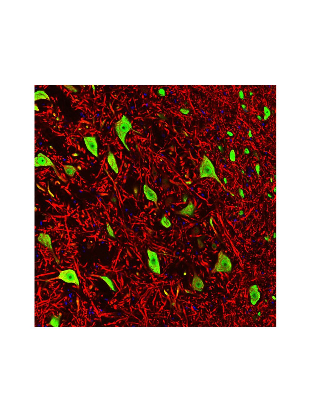

Neuronal nuclei antigen [NeuN]/FOX3, (NeuN/FOX3), Mouse Monoclonal Antibody (1B7)
- Product Name Neuronal nuclei antigen [NeuN]/FOX3, (NeuN/FOX3), Mouse Monoclonal Antibody (1B7)
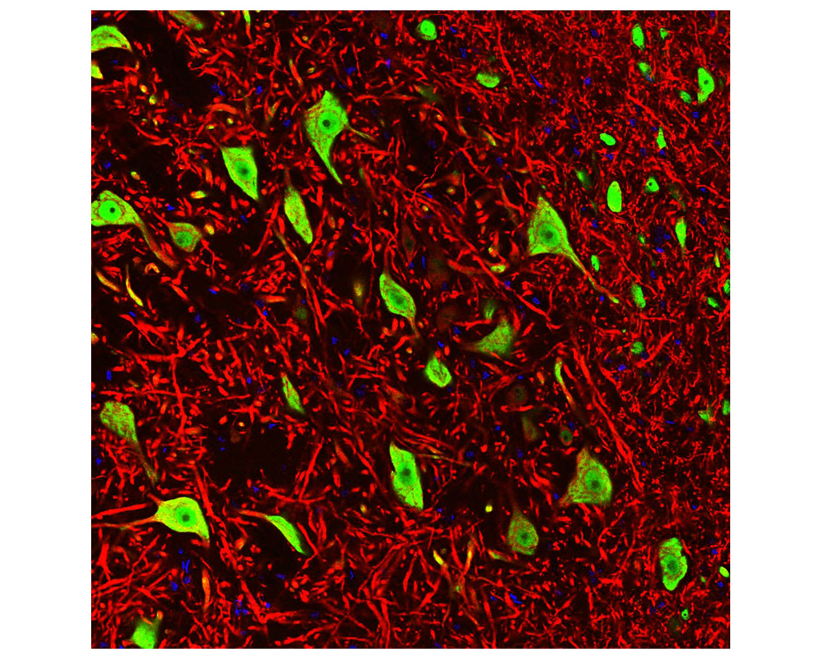
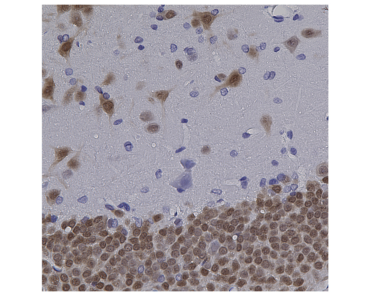
- Product Description Mouse anti-Neuronal nuclei antigen [NeuN] /RNA binding protein fox-1 homolog 3 [FOX3], (NeuN/FOX3), Monoclonal Antibody (Unconjugated), Clone 1B7, suitable for WB and Immunostaining.
- Alternative Names Neuronal nuclei antigen, NeuN, NA binding protein fox-1 homolog 3, Fox3, NeuN, A6NFN3, RBFOX3, Fox-1 homolog C
- Application(s) IF, ICC, IHC, WB
- Antibody Host Mouse
- Antibody Type Monoclonal
- Specificity Species cross-reactivity includes human, rat, and mouse.
- Species Reactivity Human, Mouse, Rat
- Immunogen Description The antibody has been made against a recombinant construct, based on the N-terminal (100 amino acids) of human FOX3 expressed in and purified from E Coli.
- Conjugate Unconjugated
- Purity Description Protein G purified
- Regulatory Status For research use only.
Product Info
- Product Description Mouse anti-Neuronal nuclei antigen [NeuN] /RNA binding protein fox-1 homolog 3 [FOX3], (NeuN/FOX3), Monoclonal Antibody (Unconjugated), Clone 1B7, suitable for WB and Immunostaining.
-
Related Products
Neuronal nuclei antigen [NeuN]/FOX3, (NeuN/FOX3), Chicken Polyclonal Antibody
Neuronal nuclei antigen [NeuN]/FOX3, (NeuN/FOX3), Rabbit Polyclonal Antibody
- Application(s) IF, ICC, IHC, WB
- Application Details Western blot (WB), Immunocytochemistry (ICC) / Immunofluorescence (IF), Immunohistochemistry (IHC). A dilution of 1:1,000 is recommended for WB. A dilution of 1:1,000 – 1:2,000 is recommended for ICC/IF and IHC. Biosensis recommends optimal dilutions/concentrations should be determined by the end user.
- Target Neuronal nuclei antigen [NeuN] / RNA binding protein fox-1 homolog 3 [FOX3] (NeuN/FOX3)
- Specificity Species cross-reactivity includes human, rat, and mouse.
- Target Host Species Human
- Species Reactivity Human, Mouse, Rat
- Antibody Host Mouse
- Antibody Type Monoclonal
- Antibody Isotype IgG2b, Κ
- Clone Name 1B7
- Conjugate Unconjugated
- Immunogen Description The antibody has been made against a recombinant construct, based on the N-terminal (100 amino acids) of human FOX3 expressed in and purified from E Coli.
- Sequence MAQPYPPAQYPPPPQNGIPAEYAPPPPHPTQDYSGQTPVPTEHGMTLYTPAQTHPEQPGSEASTQPIAGTQTVPQTDEAAQTDSQPLHPSDPTEKQQPKR
- Purity Description Protein G purified
- Format Lyophilized from PBS buffer pH 7.2-7.6 with 0.1% trehalose, and sodium azide
- Reconstitution Instructions Spin vial briefly before opening. Reconstitute with 100 µL sterile-filtered, ultrapure water to achieve a 1 mg/mL concentration. Centrifuge to remove any insoluble material.
- Storage Instructions Store lyophilized antibody at 2-8°C After reconstitution of lyophilized antibody, aliquot and store at -20°C for a higher stability. Avoid freeze-thaw cycles. Store at 4°C for up to one month for short term storage and frequent use.
- Batch Number Please see item label.
- Expiration Date 12 months after date of receipt (unopened vial).
- Alternative Names Neuronal nuclei antigen, NeuN, NA binding protein fox-1 homolog 3, Fox3, NeuN, A6NFN3, RBFOX3, Fox-1 homolog C
- Uniprot Number A6NFN3
- Uniprot Number/Name A6NFN3 (RFOX3_HUMAN)
- Scientific Background FOX3 is one of a family of mammalian homologues of FOX1 (FOX is an acronym of "Feminizing locus on X”). The FOX proteins are about 46 kDa in size, and each includes a central highly conserved RRM type RNA recognition motif. Much interest has focused on FOX3 as a result of the recent finding that this protein corresponds to NeuN, a neuronal nuclear antigen. NeuN/FOX3 functions as a Pre-mRNA alternative splicing regulator. It regulates alternative splicing of RBFOX2 to enhance the production of mRNA species that are targeted for nonsense-mediated decay (NMD) and is expressed heavily and specifically in neuronal nuclei and cytoplasm. In the cerebellum, Fox3 does not stain Purkinje neurons or Golgi neurons. The antibody was raised against the N-terminal (100 amino acids) of human FOX3, as expressed in and purified from E. coli. The full length FOX3 is not used as the immunogen since the three mammalian FOX homologues, namely FOX1, FOX2 and FOX3, include virtually identical RRM motifs. The N-terminal region of the three molecules are much more variable in the three molecules, so antibodies specific for each of the three molecules can therefore be generated.
- Shipping Temperature 25°C (ambient)
- UNSPSC CODE 41116161
- Regulatory Status For research use only.
Specifications


-
Specific References
Suzuki Y et al. (2022) "High-Throughput Screening Assay Identifies Berberine and Mubritinibas Neuroprotection Drugs for Spinal Cord Injury via Blood‑Spinal Cord Barrier Protection." Neurotherapeutics. [Epub ahead of print] Application: IHC (IF). Species: Mouse.
Yeh SJ et al. (2021) "Capping Protein Regulator and Myosin 1 Linker 3 (CARMIL3) as a Molecular Signature of Ischemic Neurons in the DWI-T2 Mismatch Areas After Stroke." Front Mol Neurosci. 14:754762. Application: IHC (IF). Species: Rat.
Han Y & Zhou XF (2019) "Method of Producing Multipotent Stem Cells." US Patent US 10,196,606 B2. Application: ICC (IF). Species: Human.
Santos J et al. (2017) "Proteomic Analysis of Human Adipose Derived Stem Cells during Small Molecule Chemical Stimulated Pre-neuronal Differentiation." Int J Stem Cells. 2017; 10(2):193-217. Application: WB. Species: Human.
Hamanoue M et al. (2016) "Cell-permeable p38 MAP kinase promotes migration of adult neural stem/progenitor cells" Sci Rep. 6:24279. Application: WB. Species: Mouse.
Han YC et al. (2016) "Direct Reprogramming of Mouse Fibroblasts to Neural Stem Cells by Small Molecules" Stem Cells Int. 2016; 2016:4304916. Application: ICC (IF). Species: Mouse.