1800 605-5127
 +61 (0)8 8352 7711
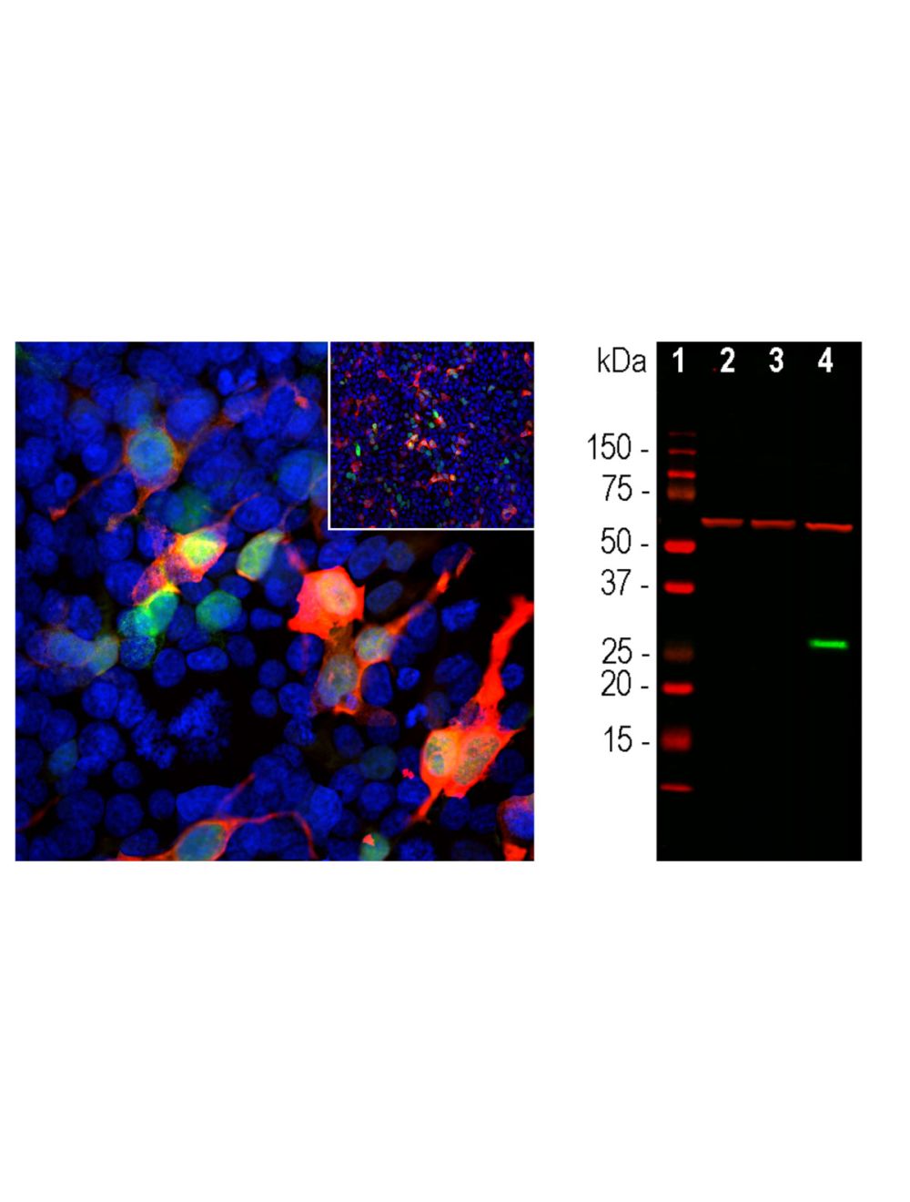

Green Fluorescent Protein Tag (GFP), Mouse Monoclonal Antibody
- Product Name Green Fluorescent Protein Tag (GFP), Mouse Monoclonal Antibody
- Product Description Mouse anti-Green fluorescent protein (GFP) Monoclonal Antibody (Unconjugated), suitable for WB, ICC.
- Alternative Names Green fluorescent protein, GFP
- Application(s) ICC, WB
- Antibody Host Mouse
- Antibody Type Monoclonal
- Specificity Specific for GFP, does not cross-react with mCherry.
- Species Reactivity Species Independent
- Immunogen Description Recombinant AcGFP protein expressed in and purified from E. Coli.
- Conjugate Unconjugated
- Purity Description Protein L purified
- Regulatory Status For research use only.
Product Info
- Product Description Mouse anti-Green fluorescent protein (GFP) Monoclonal Antibody (Unconjugated), suitable for WB, ICC.
-
Related Products
Green Fluorescent Protein Tag (GFP), Chicken Polyclonal Antibody
Green Fluorescent Protein Tag (GFP), Goat Polyclonal Antibody
Enhanced green fluorescent Purified Protein (EGFP), Purified Recombinant Protein
Green fluorescent protein (GFP), Rabbit Polyclonal Antibody
- Application(s) ICC, WB
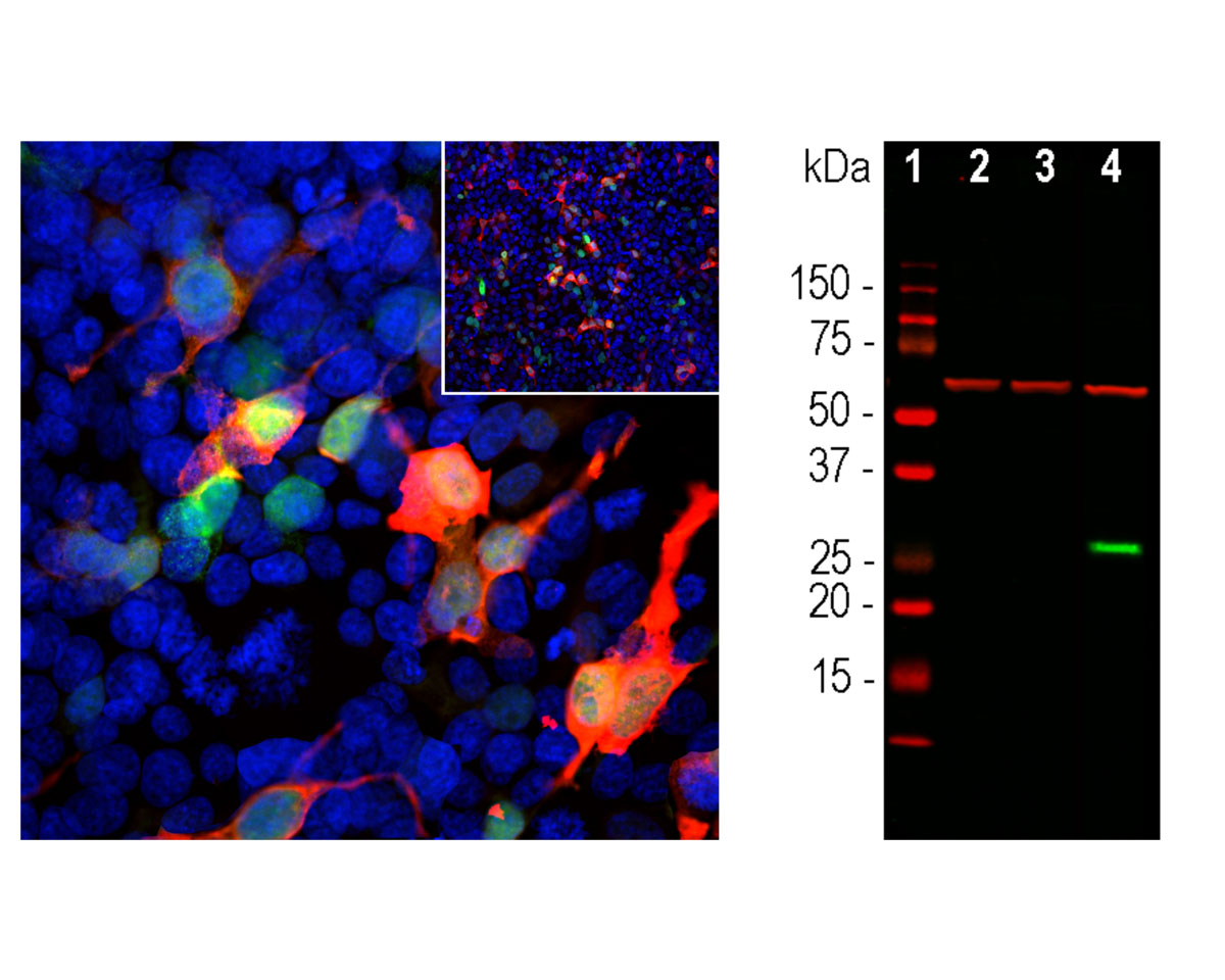
- Application Details Western blotting (1:1,000) and Immunocytochemistry (1:1,000). Biosensis recommends optimal dilutions/concentrations should be determined by the end user.
- Product Validation Antibody recognizes GFP protein in GFP-transfected HEK293 cells, but not in non-transfected control cells.
- Target Green fluorescent protein (GFP)
- Specificity Specific for GFP, does not cross-react with mCherry.
- Target Host Species Jellyfish
- Species Reactivity Species Independent
- Antibody Host Mouse
- Antibody Type Monoclonal
- Antibody Isotype IgM
- Clone Name 1F1
- Conjugate Unconjugated
- Immunogen Description Recombinant AcGFP protein expressed in and purified from E. Coli.
- Immunogen Length Full-length recombinant protein.
- Epitope The epitope is in the N-terminal 18 amino acids of the protein (peptide MVSKGAELFTGIVPILIE), which is found in the Clontech and other GFP vectors
- Positive Control GFP-transfected HEK293 cells.
- Negative Control Non-transfected HEK293 cells.
- Purity Description Protein L purified
- Physical State Solid.
- Format Lyophilized from PBS buffer pH 7.2-7.6 with 0.1% trehalose, and sodium azide
- Reconstitution Instructions Spin vial briefly before opening. Reconstitute with 100 µL sterile-filtered, ultrapure water to achieve a 1 mg/mL concentration. Centrifuge to remove any insoluble material.
- Storage Instructions Store lyophilized antibody at 2-8°C. After reconstitution divide into aliquots and store at -20°C for long-term storage. Store at 2-8°C short-term (up to 4 weeks). Avoid repetitive freeze/thaw cycles.
- Batch Number Please see item label.
- Expiration Date 12 months after date of receipt (unopened vial).
- Alternative Names Green fluorescent protein, GFP
- Uniprot Number Q6YGZ0
- Uniprot Number/Name Q6YGZ0 (Q6YGZ0_9CNID)
- Cellular Localization Intracellular, cytosolic.
-
Scientific Background
The green fluorescent protein (GFP) is a 27 kDa protein isolated originally from the jellyfish Aequoria victoria. It has an endogenous fluorochrome activity with excitation maximum at 395 nm and emission maximum at 509 nm, which is similar to that of fluorescein. GFP can be expressed in fluorescent form in essentially any prokaryotic or eukaryotic cell.
This GFP rabbit antibody was made against a recombinant GFP construct originating from an Aequoria species which was engineered to improve spectral properties and prevent oligomerization. This form of GFP, referred to as AcGFP, is 94% identical to the eGFP developed by Tsien and co-workers. The antibody can be used to verify the expression, size and stability of both AcGFP and eGFP fusion proteins in western blotting experiments and to amplify GFP signals in tissues of transgenic animals. - Shipping Temperature 25°C (ambient)
- UNSPSC CODE 41116161
- Regulatory Status For research use only.