 1800 605-5127
 +61 (0)8 8352 7711
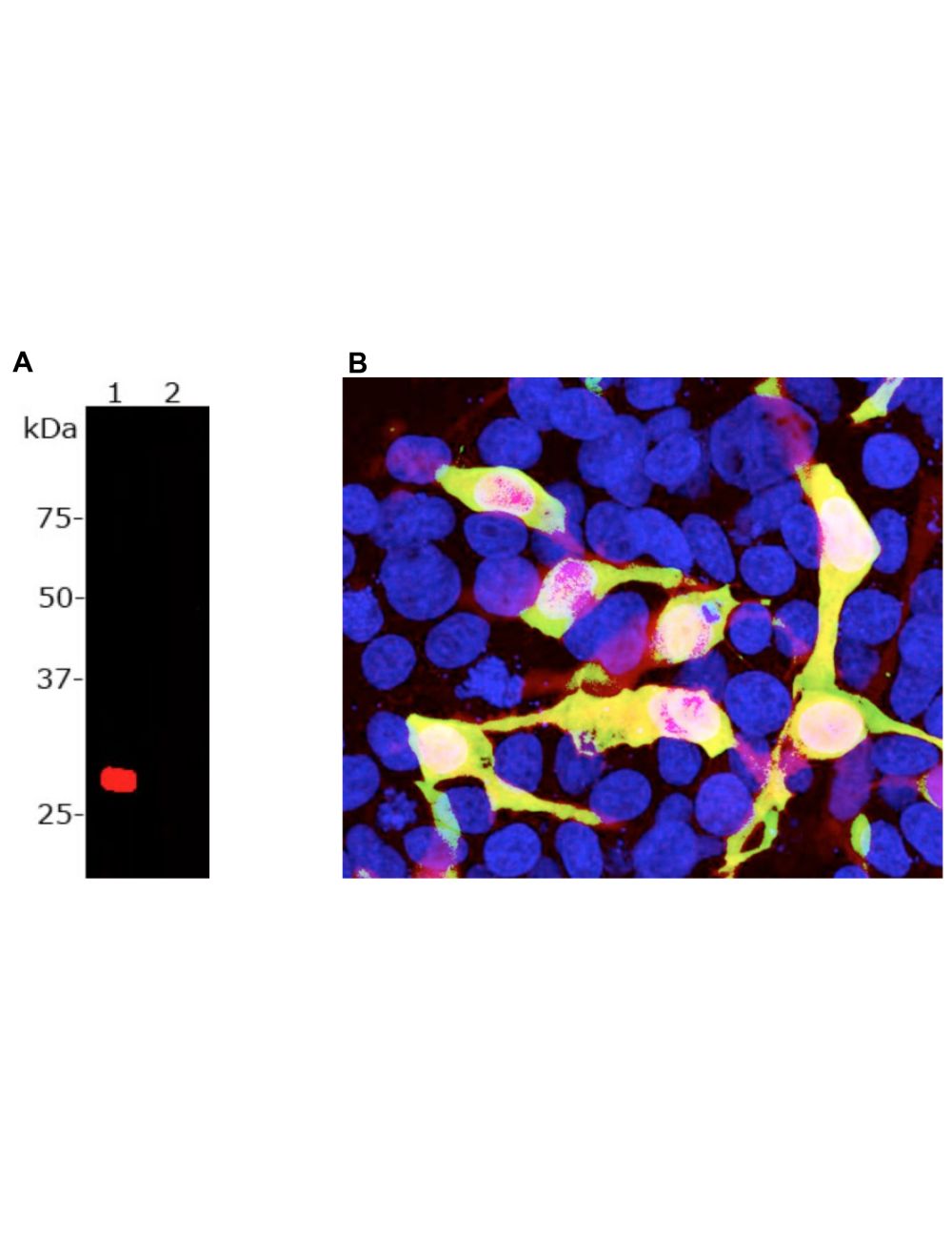

mCherry, Chicken Polyclonal Antibody
- Product Name mCherry, Chicken Polyclonal Antibody
- Product Description Chicken anti-mCherry Polyclonal Antibody (Unconjugated), suitable for WB, ICC.
- Application(s) ICC, WB
- Antibody Host Chicken
- Antibody Type Polyclonal
- Specificity The antibody reacts with a band at ~28-30 kDa corresponding to intact full-length mCherry by Western blot on HEK293 cells transfected with mCherry vector. It has also been used successfully for immunocytochemistry.
- Species Reactivity Species Independent
- Immunogen Description Recombinant full length mCherry.
- Conjugate Unconjugated
- Purity Description Concentrated purified IgY
- Regulatory Status For research use only.
Product Info
- Product Description Chicken anti-mCherry Polyclonal Antibody (Unconjugated), suitable for WB, ICC.
- Application(s) ICC, WB
- Application Details Western Blotting (WB) and Immunocytochemistry (ICC). A dilution of 1:2,000 - 1:5,000 isC recommended for WB. A dilution of 1:500-1,000 is recommended for IC. Biosensis recommends optimal dilutions/concentrations should be determined by the end user.
- Target mCherry
- Specificity The antibody reacts with a band at ~28-30 kDa corresponding to intact full-length mCherry by Western blot on HEK293 cells transfected with mCherry vector. It has also been used successfully for immunocytochemistry.
- Target Host Species Species Independent
- Species Reactivity Species Independent
- Antibody Host Chicken
- Antibody Type Polyclonal
- Antibody Isotype IgY
- Conjugate Unconjugated
- Immunogen Description Recombinant full length mCherry.
- Purity Description Concentrated purified IgY
- Format Lyophilized IgY preparation, with sodium azide.
- Reconstitution Instructions Spin vial briefly before opening. Reconstitute with 100 µL sterile-filtered, ultrapure water. Centrifuge to remove any insoluble material.
- Storage Instructions Store lyophilized, unopened vial at 2-8°C or lower. After reconstitution, prepare aliquots and store at -20°C to -80°C for a higher stability. Avoid freeze-thaw cycles.
- Batch Number Please see item label.
- Expiration Date 12 months after date of receipt (unopened vial).
- Scientific Background mCherry is an engineered derivative of one of a family of proteins originally isolated from Cnidarians (jelly fish, sea anemones and corals). The mCherry protein was derived from DsRed, a red fluorescent protein from so-called disc corals of the genus Discosoma. DsRed is a 223 amino acid ~28 kDa protein similar in size and properties to GFP, but, obviously, produces a red rather than a green fluorochrome. The original DsRed was engineered extensively in the Tsien lab to prevent it from forming tetramers and dimers and to modify and improve the spectral properties (1-3). The resulting monomeric protein is useful for applications such as Foerster Resonance Energy Transfer (FRET, also known as Fluorescence Resonance Energy Transfer). Several further cycles of mutation, directed modification and evolutionary selection produced mCherry, which is monomeric and has an excitation maximum at 587 nm and and emission maximum at 610 nm (4).
- Shipping Temperature 25°C (ambient)
- UNSPSC CODE 41116161
- Regulatory Status For research use only.
Specifications


-
General References
Baird GS, Zacharias DA, Tsien RY. Biochemistry, mutagenesis, and oligomerization of DsRed, a red fluorescent protein from coral. Proc Natl Acad Sci U S A. 97:11984-9 (2000).
Gross LA, Baird GS, Hoffman RC, Baldridge KK, Tsien RY. The structure of the chromophore within DsRed, a red fluorescent protein from coral. Proc Natl Acad Sci U S A. 97:11990-5 (2000).
Heikal AA, Hess ST, Baird GS, Tsien RY, Webb WW. Molecular spectroscopy and dynamics of intrinsically fluorescent proteins: coral red (dsRed) and yellow (Citrine). Proc Natl Acad Sci U S A. 97:11996-2001 (2000).
Shaner NC, Campbell RE, Steinbach PA, Giepmans BN, Palmer AE, Tsien RY. Improved monomeric red, orange and yellow fluorescent proteins derived from Discosoma sp. red fluorescent protein. Nature Biotechnology 22:1567-1572 (2004).