 1800 605-5127
 +61 (0)8 8352 7711
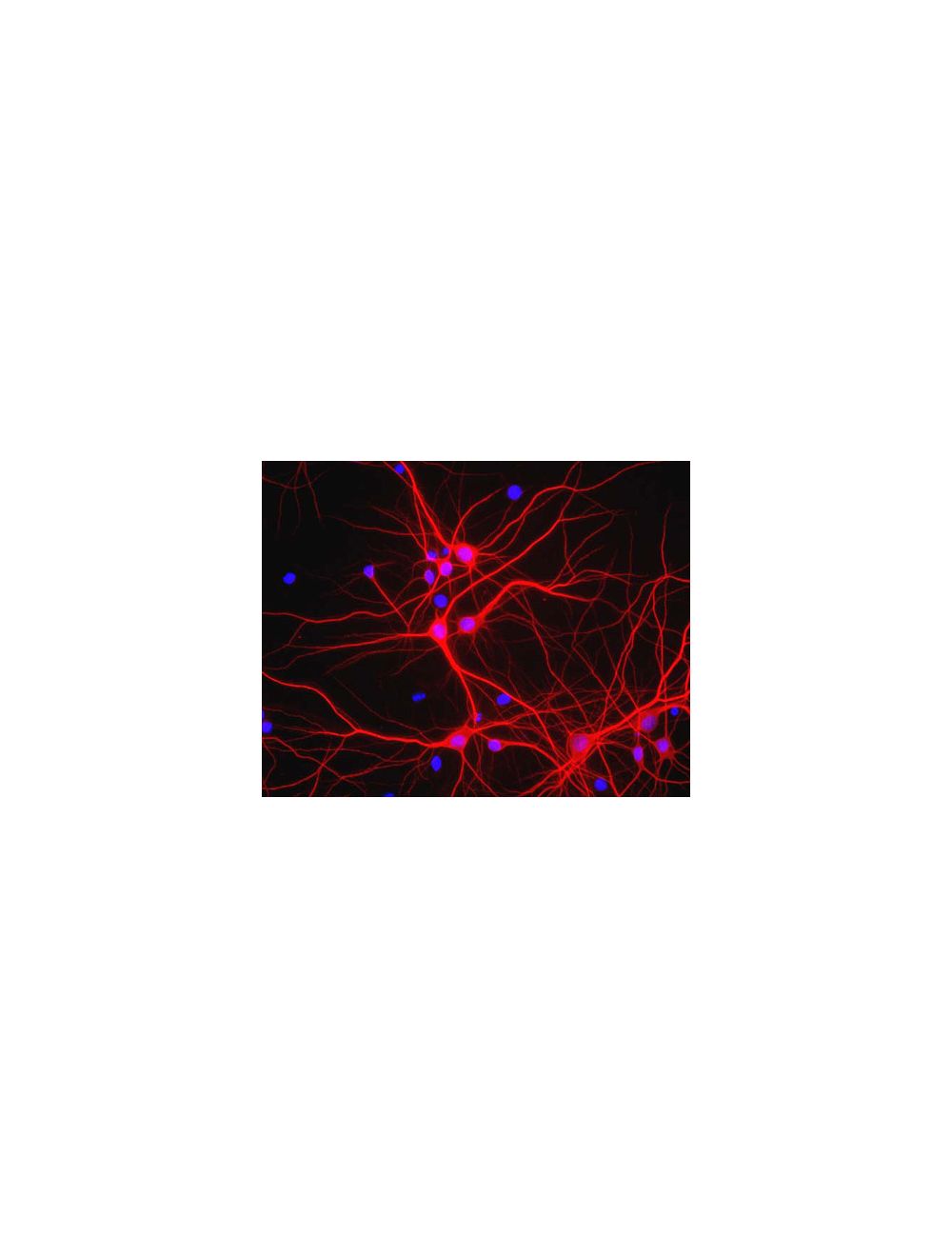

Microtubule-associated protein 2 (MAP2), Chicken Polyclonal Antibody
- Product Name Microtubule-associated protein 2 (MAP2), Chicken Polyclonal Antibody
- Product Description Chicken anti-Microtubule-associated protein 2 (MAP2) Polyclonal Antibody (Unconjugated), suitable for WB, IHC-Frozen, IHC-Paraffin-embedded, ICC.
- Alternative Names Microtubule-associated protein 2; MAP-2; MAP2;
- Application(s) ICC, IHC-Frozen, IHC-Paraffin-embedded, WB
- Antibody Host Chicken
- Antibody Type Polyclonal
- Specificity The specificity of this antibody has been confirmed by IC. Hu, Rat, Ms, Bov
- Species Reactivity Bovine, Human, Mouse, Rat
- Immunogen Description Bovine MAP2 isolated from brain by the GTP microtubule cycling method.
- Conjugate Unconjugated
- Purity Description IgY
- Regulatory Status For research use only.
Product Info
- Product Description Chicken anti-Microtubule-associated protein 2 (MAP2) Polyclonal Antibody (Unconjugated), suitable for WB, IHC-Frozen, IHC-Paraffin-embedded, ICC.
- Application(s) ICC, IHC-Frozen, IHC-Paraffin-embedded, WB
- Application Details Western Blotting (WB), Immunocytochemistry (ICC) and Immunohistochemistry (IHC). Suggested dilutions are: 1:10,00-1:20,000 (WB), 1:5,000-1:10,000 (ICC, IHC). Biosensis recommends optimal dilutions/concentrations should be determined by the end user.
- Target Microtubule-associated protein 2 (MAP2)
- Specificity The specificity of this antibody has been confirmed by IC. Hu, Rat, Ms, Bov
- Target Host Species Bovine
- Species Reactivity Bovine, Human, Mouse, Rat
- Antibody Host Chicken
- Antibody Type Polyclonal
- Antibody Isotype IgY
- Conjugate Unconjugated
- Immunogen Description Bovine MAP2 isolated from brain by the GTP microtubule cycling method.
- Purity Description IgY
- Format Lyophilized IgY preparation, with sodium azide.
- Reconstitution Instructions Spin vial briefly before opening. Reconstitute with 50 µL sterile-filtered, ultrapure water. Centrifuge to remove any insoluble material.
- Storage Instructions After reconstitution of lyophilized antibody, aliquot and store at -20°C for a higher stability. Avoid freeze-thaw cycles.
- Batch Number Please see item label.
- Expiration Date 12 months after date of receipt (unopened vial).
- Alternative Names Microtubule-associated protein 2; MAP-2; MAP2;
- Uniprot Number P11137
- Uniprot Number/Name P11137 (MTAP2_HUMAN)
- Scientific Background Microtubules are 25nm diameter protein rods found in most kinds of eukarytic cells. They are polymerized from a dimeric subunit made of one a subunit and one b tubulin subunit. Microtubules are associated with a family of proteins called microtubule associated proteins (MAPs), which includes the protein t (tau) and a group of proteins referred to as MAP1, MAP2, MAP3, MAP4 and MAP5. MAP2 is made up of two ~280 kDa apparent molecular weight bands referred to as MAP2a and MAP2b. A third lower molecular weight form, usually called MAP2c, corresponds to a pair of protein bands running at ~70 kDa on SDS-PAGE gels. All these MAP2 forms are derived from a single gene by alternate transcription, and all share a C-terminal sequence which includes either three or four microtubule binding peptide sequences, which are very similar to those found in the related microtubule binding protein t (tau). MAP2 isoforms are expressed only in neuronal cells and specifically in the perikarya and dendrites of these cells. Antibodies to MAP2 are therefore excellent markers on neuronal cells, their perikarya and neuronal dendrites.
- Shipping Temperature 25°C (ambient)
- UNSPSC CODE 41116161
- Regulatory Status For research use only.
Specifications


-
Specific References
Michurina A (2021) "Loss of Setd1b methyltransferase in the murine forebrain as a novel model for human intellectual disability." PhD Thesis. Application: IHC (IF) Species: Mouse.
Iwata M et al. (2019) "Regulatory mechanisms for the axonal localization of tau protein in neurons." Mol Biol Cell. 30(19):2441-57. Application: ICC/IF Species: Mouse, rat.
Duda JK et al. (2019) "The role of DLG-MAGUKs in mediating signaling specificity at the postsynaptic density." PhD Thesis. [Epub ahead of print]. Application: ICC/IF Species: Mouse.
Awashti A et al. (2018) "Synaptotagmin-3 drives AMPA receptor endocytosis, depression of synapse strength, and forgetting." Science. 2018; [Epub ahead of print]. Application: IHC/ICC/IF Species: Mouse.
Wolfes AC et al. (2016) "A novel method for culturing stellate astrocytes reveals spatially distinct Ca2+ signaling and vesicle recycling in astrocytic processes." J Gen Physiol. 2016; [Epub ahead of print]. Application: IF Species: Rat.
Dziennis S et al. (2007) "Role of signal transducer and activator of transcription-3 in estradiol-mediated neuroprotection." J Neurosci. 2007; 27(27):7268-74. Application: IHC Species: Rat.