 1800 605-5127
 +61 (0)8 8352 7711
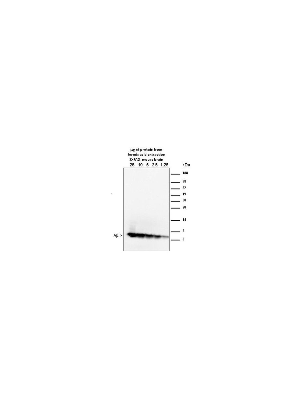

Amyloid beta peptide (A-beta 40/42), Mouse Monoclonal Antibody
- Product Name Amyloid beta peptide (A-beta 40/42), Mouse Monoclonal Antibody
-
Product Description
Amyloid beta peptide (A-beta 40/42), Mouse Monoclonal Antibody (Unconjugated), suitable for WB, IHC-Frozen, IHC-Paraffin-embedded, ICC, IP, FC, ELISA.
- Alternative Names Beta-APP42; Beta-APP40; Beta-amyloid protein 42; Beta-amyloid protein 40; ABPP; APPI; Amyloid beta A4 protein;MOAB2;MOAB-2; Alzheimer's antibody;AB40;AB42;abeta
- Application(s) ELISA, FC, ICC, IHC-Frozen, IHC-Paraffin-embedded, IP, WB
- Antibody Host Mouse
- Antibody Type Monoclonal
- Specificity MOAB-2 detects preparations enriched in U-, O-, F-Aβ42, and U-Aβ40 by dot-blot, and is thus a pan-specific Aβ antibody. However, MOAB-2 is selective for the more neurotoxic Aβ42 compared to Aβ40. Indeed, MOAB-2 demonstrated a titration against antigen concentration, and detects Aβ40 at 2.5 pmol but U-, O- and FAβb42 at antigen concentrations as low as ~ 0.1 pmol {Youmans. KL et al 2012}. MOAB-2 does not detect APP (Amyloid precursor protein). Human, Rat, other species not yet tested.By Dot blot, MOAB-2 detected rat Aβ40 and human Aβ40, albeit with less affinity than for Aβ42. {Youmans. KL et al 2012}
- Species Reactivity Human, Rat
- Immunogen Description Recombinant human amyloid beta protein 42 (Aβ42): DAEFRHDSGYEVHHQKLVFFAEDVGSNKGAIIGLMVGGVVIA
- Conjugate Unconjugated
- Purity Description This product is a Protein A purified mouse IgG2b in 0.02 M Potassium Phosphate, 0.15 M Sodium Chloride, 0.01% sodium azide, pH 7.2.
- Regulatory Status For research use only.
Product Info
-
Product Description
Amyloid beta peptide (A-beta 40/42), Mouse Monoclonal Antibody (Unconjugated), suitable for WB, IHC-Frozen, IHC-Paraffin-embedded, ICC, IP, FC, ELISA.
-
Related Products
Oligomeric Amyloid-beta, Human, Rat, ELISA assay
Amyloid beta peptide (A-beta 40/42), Mouse Monoclonal Antibody (Biotin)
- Application(s) ELISA, FC, ICC, IHC-Frozen, IHC-Paraffin-embedded, IP, WB
-
Application Details
Western Blotting (WB), Immunohistochemistry (IHC), Immunohistochemistry/paraffin embedded IH(P), Immunoprecipitation (IP), Immunofluorescence (IF), ELISA.
Antibody has been tested in WB using purified synthetic beta-amyloid preparations and from transgenic mouse brain formic acid extracts (see figure 1). Formic acid extraction/concentration is required for western blot detection from extracts. MOAB-2 antibody is specific for beta-amyloid and does not detect APP. Suggested dilution of 1:2000-1:5,000 for WB, standard ECL detection systems.
Tissue samples for the detection of beta-amyloid should be prepared as detailed in K.L. Youmans et al. {Journal of Neuroscience Methods 196 (2011) 51-59} for best results. Detection of beta-amyloid 40/42 in direct westerns can be difficult; Dot-blots of prepared samples are recommended as detailed in Youmans. KL et al 2012.
IR or fluorescent detection systems not yet tested, they but are expected to work well with higher primary antibody dilutions because of the increased sensitivity of the detection methods.
Suggested dilutions for IHC are 1:50-1:1,000. Fresh frozen, 4% paraformaldehyde fixed frozen, or formalin fixed paraffin embedded tissues are all suitable. Optimal dilutions must be determined by the end user. Antigen retrieval is required in fixed tissues for optimal staining.
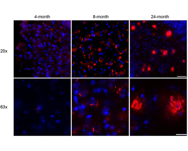
Antibody was tested on 4% paraformaldehyde/0.1% glutaraldehyde fixed frozen tissue from 3xTg and 5xFAD mice. MOAB-2 antibody detects intraneuronal and extracellular beta-amyloid in IHC and does not detect APP {Youmans KL et al 2012}.
The antibody also reacts with archival formalin-fixed, paraffin-embedded tissue samples with antigen Heat Induced Epitope Retrieval (HIER): Recommended Citrate, pH 6.0 buffer for HIER. Signal was weak without antigen retrieval. Immunoreactively was expressed in intraneural-amyloid deposition (plaque) in Alzheimer's brain. MoAB-2 was found to be extremely clean and with an excellent signal to noise ratio with no neuro-cellular diffusive staining.
In addition MOAB-2 demonstrated no significant differences in A-beta detection using paraffin fixed, free-floating sections {Youmans KL et al 2012}. Formic acid (FA) treatment resulted in optimal detection of both intraneuronal and extracellular A-beta compared to without FA (incubated in 88% FA 8 min, Youmans KL et al 2012). Free floating tissue sections were permeabilized in TBS containing 0.25% Triton X-100 (TBSX; 3 x 10 min), blocked with 3% horse serum in TBSX (3 x 10 min) followed by 1% horse serum in TBSX (2 x10 min) and incubated with appropriate primary antibodies diluted in TBSX containing 1% horse serum overnight. See Youmans KL et al 2012 for full IH(P) protocol and method details.
For IF, suggested dilution is 1:100-1:500. The antibody was tested on 4% PFA fixed frozen tissue. Fixed tissues were washed in TBS (3 x 10 min), then incubated in 88% FA (8 min), and then permeabilized in TBSX (3 x 10 min), and blocked in TBSX containing 5% bovine serum albumin (BSA; 1 hr). Sections were subsequently incubated with appropriate primary antibodies diluted in TBSX containing 2% BSA overnight on an oscillatory rotator. Detection was via fluorescently labelled absorbed secondary antibodies {Youmans KL et al 2012}.
For IP, the suggested dilution is 1:200 to 1:1,000 for labeled beta-amyloid using Protein A/G conjugated beads as the capture vehicle {Youmans KL et al 2012}.
In an ELISA, a dilution of 1:50-1:1000 is suggested. The antibody has been tested in ELISAs on synthetic beta-amyloid and tissue homogenates from beta-amyloid-Tg mice. Biosensis recommends optimal dilutions/concentrations should be determined by the end user for all applications. Dilutions provided are only meant to serve as a basic guide. - Target Amyloid beta peptide (A-beta 40/42)
- Specificity MOAB-2 detects preparations enriched in U-, O-, F-Aβ42, and U-Aβ40 by dot-blot, and is thus a pan-specific Aβ antibody. However, MOAB-2 is selective for the more neurotoxic Aβ42 compared to Aβ40. Indeed, MOAB-2 demonstrated a titration against antigen concentration, and detects Aβ40 at 2.5 pmol but U-, O- and FAβb42 at antigen concentrations as low as ~ 0.1 pmol {Youmans. KL et al 2012}. MOAB-2 does not detect APP (Amyloid precursor protein). Human, Rat, other species not yet tested.By Dot blot, MOAB-2 detected rat Aβ40 and human Aβ40, albeit with less affinity than for Aβ42. {Youmans. KL et al 2012}
- Target Host Species Human
- Species Reactivity Human, Rat
- Antibody Host Mouse
- Antibody Type Monoclonal
- Antibody Isotype IgG2b, lambda
- Clone Name MOAB-2
- Conjugate Unconjugated
- Immunogen Description Recombinant human amyloid beta protein 42 (Aβ42): DAEFRHDSGYEVHHQKLVFFAEDVGSNKGAIIGLMVGGVVIA
- Purity Description This product is a Protein A purified mouse IgG2b in 0.02 M Potassium Phosphate, 0.15 M Sodium Chloride, 0.01% sodium azide, pH 7.2.
- Format Lyophilized, from a Protein A purified preparation in 0.02 M Potassium Phosphate, 0.15 M Sodium Chloride, 0.01% sodium azide, 0.1% trehalose, pH 7.2; contains 0.01% sodium azide as a preservative.
- Reconstitution Instructions Spin vial briefly before opening. Reconstitute in 100 µL sterile-filtered, ultrapure water. Centrifuge to remove any insoluble material. Final buffer is 0.02 M Potassium Phosphate, 0.15 M Sodium Chloride, 0.01% sodium azide, 0.1% trehalose, pH 7.2.
- Storage Instructions After reconstitution keep aliquots at -20 ° to -70°C for a higher stability. At 2-8°C keep up to one week, insulated, protected from light; use sterile methods and pipettes. Highly purified glycerol (1:1) may be added for an additional stability. Avoid repetitive freeze/thaw cycles. Keep tightly closed when not in use and protected from light.
- Batch Number Please see item label.
- Expiration Date 12 months after date of receipt (unopened vial).
- Alternative Names Beta-APP42; Beta-APP40; Beta-amyloid protein 42; Beta-amyloid protein 40; ABPP; APPI; Amyloid beta A4 protein;MOAB2;MOAB-2; Alzheimer's antibody;AB40;AB42;abeta
- Uniprot Number P05067
- Uniprot Number/Name P05067 (A4_HUMAN)
-
Scientific Background
The amyloid beta peptide is derived from the cleavage of the Amyloid precursor protein (APP) and varies in length from 39 to 43 amino acids. However, the form(s) of amyloid-beta peptide (Aβ associated with the pathology characteristic of Alzheimer's disease (AD) remains unclear. In particular, the neurotoxicity of intraneuronal Aβ accumulation is an area of considerable research and controversy principally because antibodies thought to be specific for Aβ have been shown to actually detect intraneuronal APP and not Aβ exclusively.
MOAB-2 (mouse IgG2b) is a pan-specific, high-titer antibody to Aβ residues 1-4 as demonstrated by biochemical and immunohistochemical analyses (IHC), and is highly specific just to amyloid beta peptide.
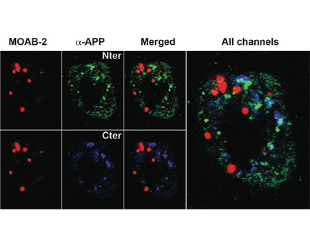
MOAB-2 did not detect APP or APP-CTFs in cell culture media/lysates (HEK-APPSwe or HEK APPSwe/BACE1) or in brain homogenates from transgenic mice expressing 5 familial AD (FAD) mutation (5xFAD mice).
Using IHC on 5xFAD brain tissue, MOAB-2 immunoreactivity co-localized with C-terminal antibodies specific for Aβ40 and Aβ42. MOAB-2 did not co-localize with either N- or C-terminal antibodies to APP. In addition, no MOAB-2-immunreactivity was observed in the brains of 5xFAD/BACE-/- mice, although significant amounts of APP were detected by N- and C-terminal antibodies to APP, as well as by 6E10.
In both 5xFAD and 3xTg mouse brain tissue, MOAB-2 co-localized with cathepsin-D, a marker for acidic organelles, further evidence for intraneuronal Aβ, distinct from Aβ associated with the cell membrane. MOAB-2 demonstrated strong intraneuronal and extra-cellular immunoreactivity in 5xFAD and 3xTg mouse brain tissues. - Shipping Temperature 25°C (ambient)
- UNSPSC CODE 41116161
- Regulatory Status For research use only.
Specifications


-
Specific References
Malik N et al. (2024) Regional AT 8 reactive tau species correlate with intracellular AB levels in cased of low AD neuropathology change Acta Neuropathology. 147(1):40 Application: Human IHC
Nuñez‑Diaz C et al. (2024) The fuorescent ligand bTVBT2 reveals increased p‑tau uptake by retinal microglia in Alzheimer’s disease patients and AppNL−F/NL−F mice Alzheimers Res Ther. 16:4 Application: IHC.
Liu J et al. (2023) Prevention of Alzheimer pathology by blocking neuregulin signaling on microglia eNeuro. [Epub ahead of print] Application: IHC.
Zhang Y et al. (2023) On-line clearing and staining method for the efficient optical imaging of large volume samples at the cellular resolution Biomed. Opt. Express. 14(9):4800-4813 Application: IHC.
Garcia MG. et al. (2023) Maternal separation differentially modulates early pathology by sex in 5xFAD Alzheimer’s disease-transgenic mice Brain Behav. Immun. [Epub ahead of print] Application: IHC.
Fang X. et al. (2023) Amyloid beta accumulation in TgF344-AD rats is associated with reduced cerebral capillary endothelial Kir2.1 expression and neurovascular uncoupling Geroscience. [Epub ahead of print] Application: IHC.
Mabrouk R. et al. (2023) Most dystrophic neurites in the common 5xFAD Alzheimer mouse model originate from axon terminals Neurobiol Dis. [Epub ahead of print] Application: IHC.
Long L. et al. (2022) Microglial hexokinase 2 deficiency increases ATP generation through lipid metabolism leading to β-amyloid clearance Nat Metab. [Epub ahead of print] Application: IHC.
Setti, S.E. et al. (2022) Assessment of sex-related neuropathology and cognitive deficits in the Tg-SwDI mouse model of Alzheimer’s disease. Behave Brain Res. 428:113882. Application: IHC.
Sil, A. et al. (2022) Sex Differences in Behavior and Molecular Pathology in the 5XFAD Model. J Alzheimers Dis. 85(2):755-778. Application: WB.
Sarkar, S. et al. (2020) Modification of methods to use Congo-red stain to simultaneously visualize amyloid plaques and tangles in human and rodent brain tissue sections. Metab Brain Dis. [Epub ahead of print]. Application: IHC.
Cuevas, E. et al. (2019) Amyloid Beta 25-35 induces blood-brain barrier disruption in vitro. Metab Brain Dis. [Epub ahead of print]. Application: ICC/IF.
Schmued, L. et al. (2019) High Contrast and Resolution Labeling of Amyloid Plaques in Tissue Sections from APP-PS1 Mice and Humans with Alzheimer's Disease with the Zinc Chelator HQ-O: Practical and Theoretical Considerations. Curr Alzheimer Res. 16(7):577-586. Application: IHC/IF.
Hui, L. et al. (2019) Acidifying Endolysosomes Prevented Low-Density Lipoprotein-Induced Amyloidogenesis. J Alzheimers Dis. 64(1):393-410. Application: ICC/IF.
Koss, DJ. et al. (2018) Distinctive temporal profiles of detergent-soluble and -insoluble tau and Aβ species in human Alzheimer's disease. Brain Res. [Epub ahead of print]. Application: WB, dot blot.
Zhao, Y. et al. (2018) TREM2 Is a Receptor for _-Amyloid that Mediates Microglial Function. Neuron. 97(5):1023-1031. Application: IHC, free-floating cryostat sections
Zhu, B. et al. (2017) ER-associated degradation regulates Alzheimer's amyloid pathology and memory function by modulating _-secretase activity. Nat Commun. 8(1):1472. Application: IHC
Huang, TY. et al. (2017) SORLA attenuates EphA4 signaling and amyloid _-induced neurodegeneration. J Exp Med. pii: jem.20171413. [Epub ahead of print]. Application: IHC
Felecia, M. et al. (2017) Peripheral Inflammation, Apolipoprotein E4, and Amyloid-_ Interact to Induce Cognitive and Cerebrovascular Dysfunction. ASN Neuro. 9(4):1759091417719201. Application: IHC/IF
Thomas, R. et al. (2016) Epidermal growth factor prevents APOE4 and amyloid-beta-induced cognitive and cerebrovascular deficits in female mice. Acta Neuropathol Commun. 4(1):111 Application: IHC
Koster, KP. et al. (2016) Epidermal growth factor prevents oligomeric amyloid-_ induced angiogenesis deficits in vitro. J Cereb Blood Flow Metab. [Epub ahead of print] Application: IF
Loffler, T. et al. (2016) Decreased Plasma Aβ in Hyperlipidemic APPSL Transgenic Mice Is Associated with BBB Dysfunction. Front. Neurosci. Application: IF
Kobro-Flatmoen, A. et al. (2016) Reelin-immunoreactive neurons in entorhinal cortex layer II selectively express intracellular amyloid in early Alzheimer's disease. Neurobiology of Disease. 93:172-183. Application: IHC
Tai, LM. et al. (2016) The role of APOE in cerebrovascular dysfunction. Acta Neuropathol. 131(5):709-23. Application: IF
Kim, YH. et al. (2015) A 3D human neural cell culture system for modeling Alzheimer's disease. Nat Prot. 10(7):985-1006. Application: WB
Condello, C. et al. (2015) Microglia constitute a barrier that prevents neurotoxic protofibrillar Aβ42 hotspots around plaques. Nat Commun. 6:6176. Application: IF
Iulita MF et al (2014) Studying Alzheimer's Disease Pre-clinical Stages: Insights from Down's Syndrome and Transgenic Animal Models. PhD Thesis Application: IHC/IF
Iulita MF et al (2014) Intracellular Abeta pathology and early cognitive impairments in a transgenic rat model overexpressing human amyloid precursor protein: a multidimensional study. Acta Neuropathol Commun. 6:61. Application: IF, IH
Smith BR et al (2014) Neuronal inclusions of alpha-synuclein contribute to the pathogenesis of Krabbe disease. J Pathol. Apr;235(5):509-21. Application: IF -
General References
Tai LM et al (2016) "The role of APOE in cerebrovascular dysfunction."Acta Neuropathol. 2016 Feb 16. [Epub ahead of print]
K.L. Youmans et al (2012) Intraneuronal Abeta detection in 5xFAD mice by a new Abeta-specific antibody Mol Neurodegener. 2012 Mar 16;7(1):8.
Tai LM et al (2013) Levels of soluble apolipoprotein E/amyloid-_ (Aβ) complex are reduced and oligomeric Aβ increased with APOE4 and Alzheimer disease in a transgenic mouse model and human samples. J Biol Chem. 2013 Feb 22;288(8):5914-26.
K.L. Youmans et al (2011) Amyloid-_42 alters apolipoprotein E solubility in brains of mice with five familial AD mutations J Neurosci Methods. 2011 Mar 15;196(1):51-9.