 1800 605-5127
 +61 (0)8 8352 7711
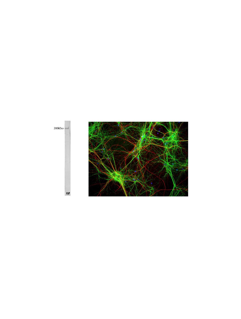

Microtubule Associated Protein 2 (MAP2), Mouse Monoclonal Antibody
- Product Name Microtubule Associated Protein 2 (MAP2), Mouse Monoclonal Antibody
- Product Description Mouse anti-Microtubule Associated Protein 2 (MAP2) Monoclonal Antibody (Unconjugated), suitable for WB, IHC-Frozen, ICC.
- Alternative Names Microtubule-associated protein 2; MAP-2; Mtap2;
- Application(s) ICC, IHC-Frozen, WB
- Antibody Host Mouse
- Antibody Type Monoclonal
- Specificity The specificity of this antibody has been confirmed by WB and IHC against the antigen. Human; Rat; Mouse;
- Species Reactivity Bovine, Human, Mouse, Rat
- Immunogen Description High molecular MAP protein preparation derived from bovine brain
- Conjugate Unconjugated
- Purity Description Protein G purified
- Regulatory Status For research use only.
Product Info
- Product Description Mouse anti-Microtubule Associated Protein 2 (MAP2) Monoclonal Antibody (Unconjugated), suitable for WB, IHC-Frozen, ICC.
- Application(s) ICC, IHC-Frozen, WB
- Application Details Immunohistochemistry (IHC), Immunocytochemistry (ICC) and Western Blotting (WB). A dilution of 1:1,000 - 1:5,000 is recommended for IHC and ICC, and 1:5,000-1:10,000 is recommended for WB. The optimal dilution should be determined by the end user.
- Target Microtubule Associated Protein 2 (MAP2)
- Specificity The specificity of this antibody has been confirmed by WB and IHC against the antigen. Human; Rat; Mouse;
- Target Host Species Bovine
- Species Reactivity Bovine, Human, Mouse, Rat
- Antibody Host Mouse
- Antibody Type Monoclonal
- Antibody Isotype IgG
- Clone Name 5H11
- Conjugate Unconjugated
- Immunogen Description High molecular MAP protein preparation derived from bovine brain
- Purity Description Protein G purified
- Format Lyophilized from PBS buffer pH 7.2-7.6 with 0.1% trehalose, and sodium azide
- Reconstitution Instructions Spin vial briefly before opening. Reconstitute with 100 µL sterile-filtered, ultrapure water to achieve a 1 mg/mL concentration. Centrifuge to remove any insoluble material.
- Storage Instructions At least 12 months after purchase at 2-8°C (lyophilized formulations). After reconstitution, aliquot and store at -20°C for a higher stability. Avoid freeze-thaw cycles.
- Batch Number Please see item label.
- Expiration Date 12 months after date of receipt (unopened vial).
- Alternative Names Microtubule-associated protein 2; MAP-2; Mtap2;
- Uniprot Number Q0IIA8
- Uniprot Number/Name Q0IIA8 (Q0IIA8_BOVIN)
- Scientific Background Microtubules are 25nm diameter protein rods found in most kinds of eukaryotic cells. They are polymerized from a dimeric subunit made of one 'a' subunit and one 'b' tubulin subunit. Microtubules are associated with a family of proteins called microtubule associated proteins (MAPs), which includes the protein t (tau) and a group of proteins referred to as MAP1, MAP2, MAP3, MAP4 and MAP5. MAP2 is made up of two ~280 kDa apparent molecular weight bands referred to as MAP2 a and MAP2 b. A third lower molecular weight form, usually called MAP2c, corresponds to a pair of protein bands running at ~70 kDa on SDS-PAGE gels. All these MAP2 forms are derived from a single gene by alternate transcription, and all share a C-terminal sequence which includes either three or four microtubule binding peptide sequences, which are very similar to those found in the related microtubule binding protein t (tau). MAP2 isoforms are expressed only in neuronal cells and specifically in the perikarya and dendrites of these cells. Antibodies to MAP2 are therefore excellent markers on neuronal cells, their perikarya and neuronal dendrites.
- Shipping Temperature 25°C (ambient)
- UNSPSC CODE 41116161
- Regulatory Status For research use only.


