Product DescriptionMouse anti-Calbindin-binding protein Monoclonal Antibody (Unconjugated), suitable for WB, IHC-Frozen.
Application(s)IHC-Frozen, WB
Application DetailsWestern blotting (1:1,000-1:5,000) and Immunohistochemistry (1:1,000). Biosensis recommends optimal dilutions/concentrations should be determined by the end user.
TargetCalbindin-binding protein
SpecificityHuman, reacts with Human, horse, Cow, Pig, chicken, Rat, Mouse.
Target Host SpeciesHuman
Species ReactivityBovine, Chicken, Horse, Human, Mouse, Pig, Rat
Antibody HostMouse
Antibody TypeMonoclonal
Antibody IsotypeIgG1
Clone Name4H7
ConjugateUnconjugated
Immunogen DescriptionFull-length recombinant human protein
Purity DescriptionProtein G purified
FormatLyophilized from PBS buffer pH 7.2-7.6 with 0.1% trehalose, and sodium azide
Reconstitution InstructionsSpin vial briefly before opening. Reconstitute with 100 µL sterile-filtered, ultrapure water to achieve a 1 mg/mL concentration. Centrifuge to remove any insoluble material.
Storage InstructionsStore lyophilized antibody at 2-8°C. After reconstitution divide into aliquots and store at -20°C for long-term storage. Store at 2-8°C short-term (up to 4 weeks) with an appropriate antibacterial agent. Avoid repetitive freeze/thaw cycles.
Batch NumberPlease see item label.
Expiration Date12 months after date of receipt (unopened vial).
Alternative NamesCalbindin D28,D-28K, Vitamin D-dependent calcium-binding protein, avian-type
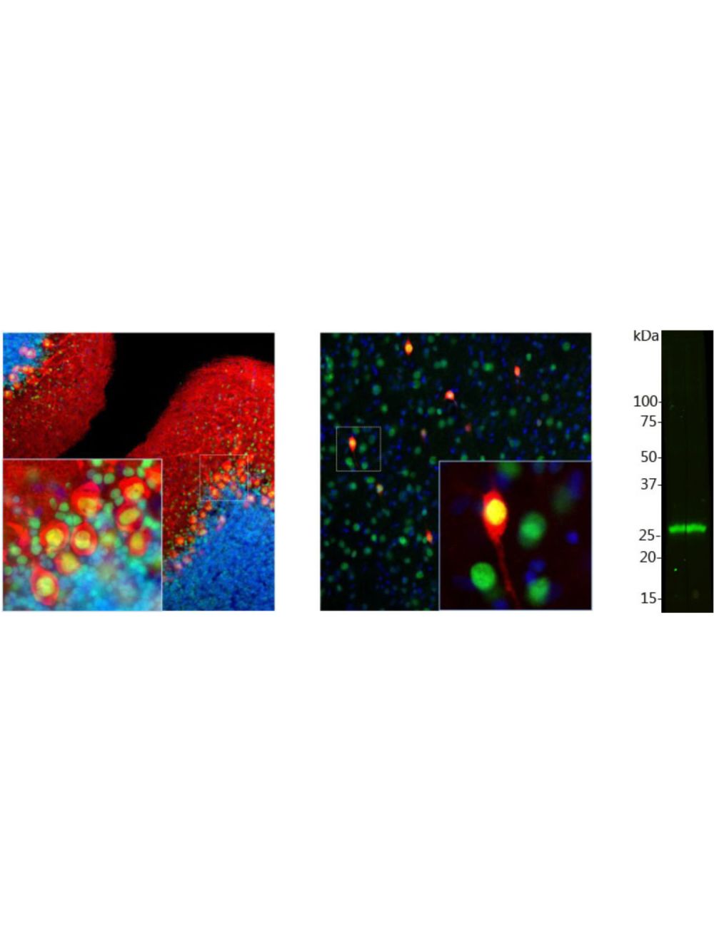
Left and middle: Detection of calbindin immunoreactivity in rat brain cerebellum (Left) and cortex (Middle) sections by Immunohistochemistry. Calbindin (red) is predominantly expressed in the dendrites and perikarya of Purkinje cells in the molecular layer of cerebellum (left), and selectively expressed in certain type of interneurons (calbindin-postive interneuron) in the cortex (middle). Methyl-CpG binding protein 2 (Rabbit anti-MeCP2, R-1810-100, green) is universally expressed in the nuclei of almost all neurons. As a result, double-immunofluorescence shows that calbindin-expressing cell somas appear red, but the nucleus appears to be yellow. Blue: DAPI nuclear stain. Insets represent high magnification images of the boxed areas. IHC method: 45 um sections, fixed by transcardial perfusion with 4% paraformaldehyde. Right: Western blot analysis of calbindin expression in cow cerebellum homogenate. The antibody shows strong immunoractivity with one calbindin band at ~30 kDa.
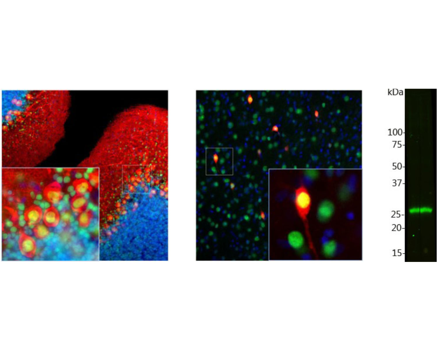
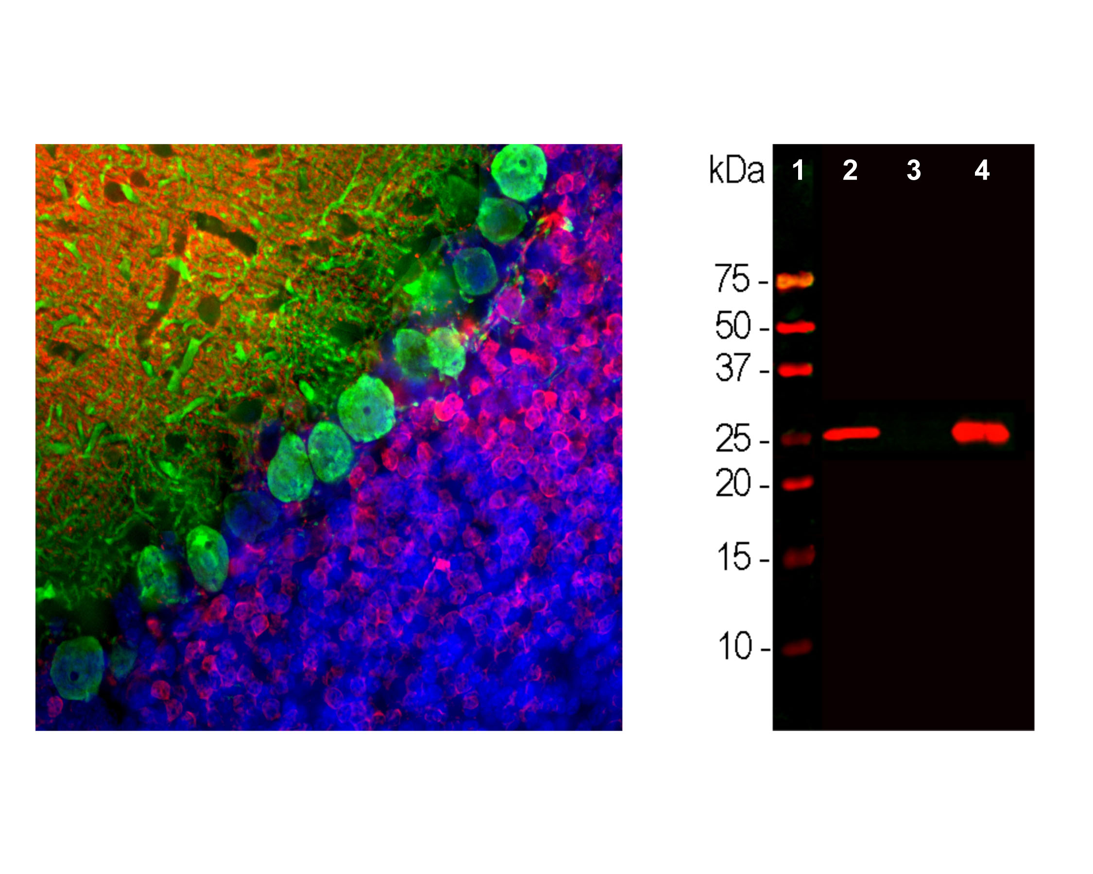
Left: Rat brain cerebellum stained with mouse anti-calbindin (green, 1:1,000) and rabbit anti-calretinin (R-1800-50, red, 1:5,000) by Immunohistochemistry. Blue: DAPI nuclear stain. IHC Method: Following transcardial perfusion with 4% paraformaldehyde, brain was post fixed for 24 hours, cut to 45 um, and free-floating sections were stained. Calbindin antibody prominently labels the dendrites and perikarya of Purkinje cells in the molecular layer of cerebellum. In contrast, the calretinin antibody stains granule cells, in the granualar layer, and their processes in the molecular layer. Right: Western blot analysis of calbindin expression (antibody dilution: 1:2,000) in rat cerebellum (2), pig hippocampus (3), and cow cerebellum (4). The band at ~25 kDa corresponds to calbindin protein, heavily expressed in the cerebellum, but less in hippocampus.
 1800 605-5127
 +61 (0)8 8352 7711