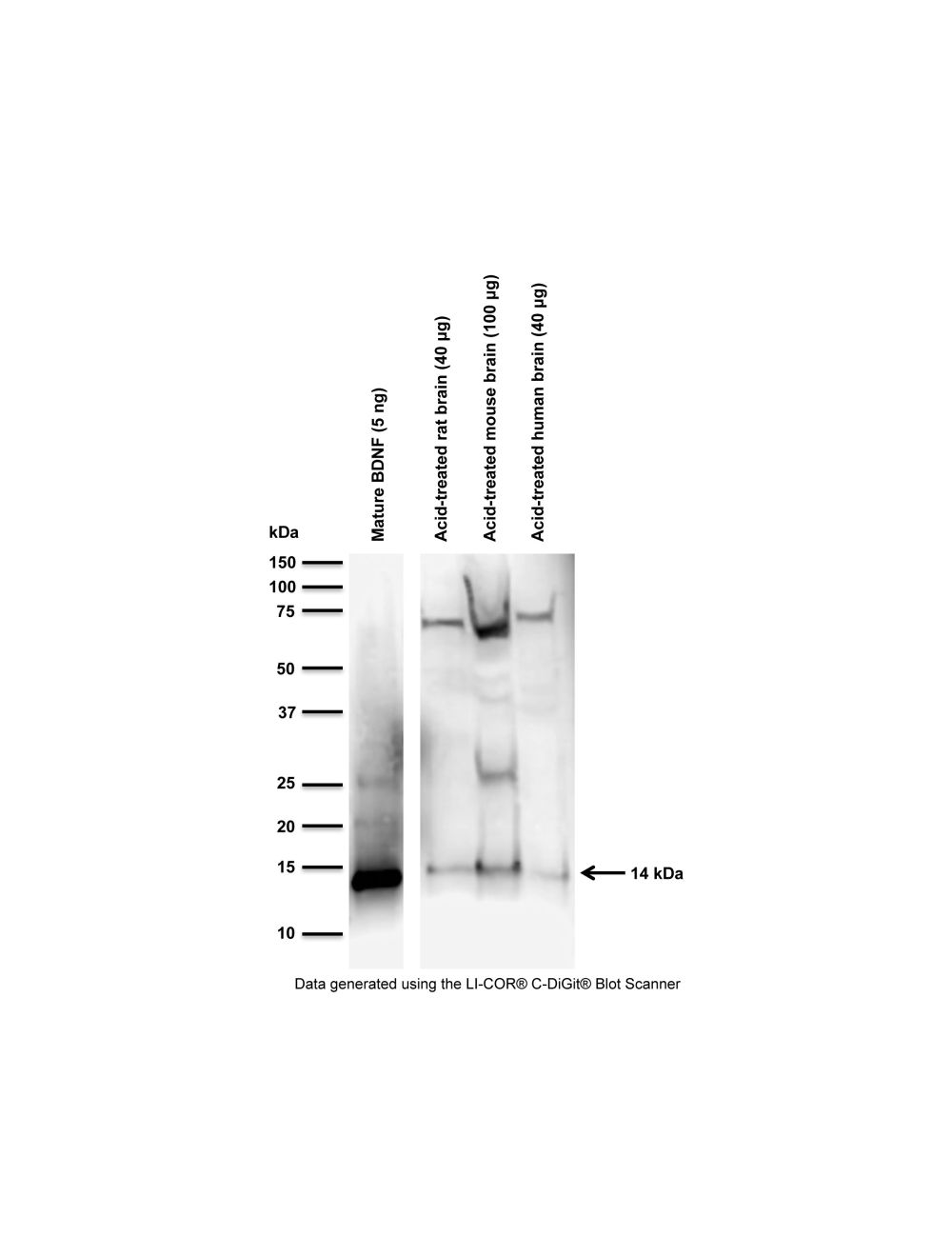
Western Blotting (denaturing and reducing): 0.2 to 2 µg/mL. M-1744-100 antibody detects 14 kDa mature BDNF monomer in human serum human and acid treated human and rodent whole tissue homogenates. In simple Tris-homogenate human brain lysates, M-1744-100 detects mature BDNF at 14kDa as well as an uncharacterized band at ~50kDa. No proBDNF band is detected in our samples, although M-1744-100 will detect proBDNF in purified form on blots. Additionally, M-1744-100 detects an additional band 18 kDa BDNF isoform human serum. For cell lysates, some caution is advised as in our hands M-1744-100 fails to detect mature BDNF on SH-SY5Y or C6 cell lysates but does detect mature BDNF in neuronal cell culture lysates. The reason for these differences has not been characterized. For detection of mature BDNF in cell lysates such as SH-SY5Y we recommend affinity-purified rabbit polyclonal antibody to rhBDNF (R-1707-100), or rabbit polyclonal antibody to BDNF peptide 1-10 (R-083-100, whole serum; R-066-500, IgG).
Immunofluorescence: 1 to 4 µg/mL, 4% formaldehyde fixation and permeabilization. Staining can be weak until optimized.
M-1744-100 is not recommended for Flow Cytometry at this time.
Biosensis recommends optimal dilutions/concentrations should be determined by the end user.
TargetBrain-derived neurotrophic factor (BDNF)
SpecificityDetects human, mouse, rat, guinea pig BDNF. Expected to detect BDNF from other species due to sequence homology.
Target Host SpeciesHuman
Species ReactivityHuman, Mouse, Other Mammals (Predicted), Rat
Antibody HostMouse
Antibody TypeMonoclonal
Antibody IsotypeIgG
Clone Name4C8
ConjugateUnconjugated
Immunogen DescriptionRecombinant human mature BDNF expressed in E.coli
Purity DescriptionProtein G purified mouse IgG.
FormatLyophilized from a solution containing PBS pH 7.4, 0.1% trehalose, with 0.1% sodium azide.
Reconstitution InstructionsSpin vial briefly before opening. Reconstitute with 50 µL sterile-filtered, ultrapure water to obtain a concentration of 1 mg/mL. Centrifuge to remove any insoluble material.
Storage InstructionsStore lyophilized antibody at -20°C to -80°C protected from moisture. After reconstitution divide antibody into useful aliquots and keep aliquots at -20°C to -80°C for a higher stability. Working aliquots can be kept at 2-8°C for up to 1 month. Avoid repetitive freeze/thaw cycles.
Batch NumberPlease see item label.
Expiration Date12 months after date of receipt (unopened vial).
Alternative NamesBrain-derived neurotrophic factor; Abrineurin; proBDNF
Scientific BackgroundBDNF belongs to the neurotrophin family and promotes the survival of neuronal populations that are all located either in the central nervous system or directly connected to it. It is a major regulator of synaptic transmission and plasticity at adult synapses in many regions of the CNS. The versatility of BDNF is emphasized by its contribution to a range of adaptive neuronal responses including long-term potentiation (LTP), long-term depression (LTD), certain forms of short-term synaptic plasticity, as well as homeostatic regulation of intrinsic neuronal excitability. The alterations in BDNF expression induced by various kinds of brain insult including stress, ischemia, seizure activity and hypoglycemia, may contribute to some pathologies such as depression, epilepsy, Alzheimer's, and Parkinson's disease. Microglia release BDNF that may contribute to neuroinflammation and neuropathic pain. SUBUNIT: Monomers and homodimers. Binds to NTRK2/TRKB. SUBCELLULAR LOCATION: Secreted protein. POst translation modification: Converted into mature BDNF by plasmin (PLG). SIMILARITY: Belongs to the NGF-beta family.
Western blot analysis of BDNF expression in acid-treated rodent and human brain samples. Brain samples were acid-treated at pH 4 (refer to References for a detailed extraction method protocol). M-1744-50/100 (1 µg/mL) detects BDNF monomer at 14 kDa. Additional higher MW bands are not characterized, but may be related to the cross-reaction of the secondary anti-mouse-HRP antibody with endogenous, reduced IgG present in samples, in particular mouse tissue.
Western Blotting Method: SDS-PAGE: denaturing and reducing, 12% Bis-Tris gel; Transfer: Tris-Glycine buffer, semi-dry transfer; Membrane: nitrocellulose (0.22 µm); Blocking: 5% skim milk in TBST, 1 hour at RT; Primary antibody: overnight at 4°C; Secondary antibody: anti-mouse-HRP (1/6000), 2 hours at RT; Detection: Chemiluminiscence.
Specific ReferencesLe Blanc J et al. (2020) Platelets Selectively Regulate the Release of BDNF, But Not That of Its Precursor Protein, proBDNF. Front Immunol. 11:575607Application: Human, WB.
 1800 605-5127
 +61 (0)8 8352 7711