 1800 605-5127
 +61 (0)8 8352 7711
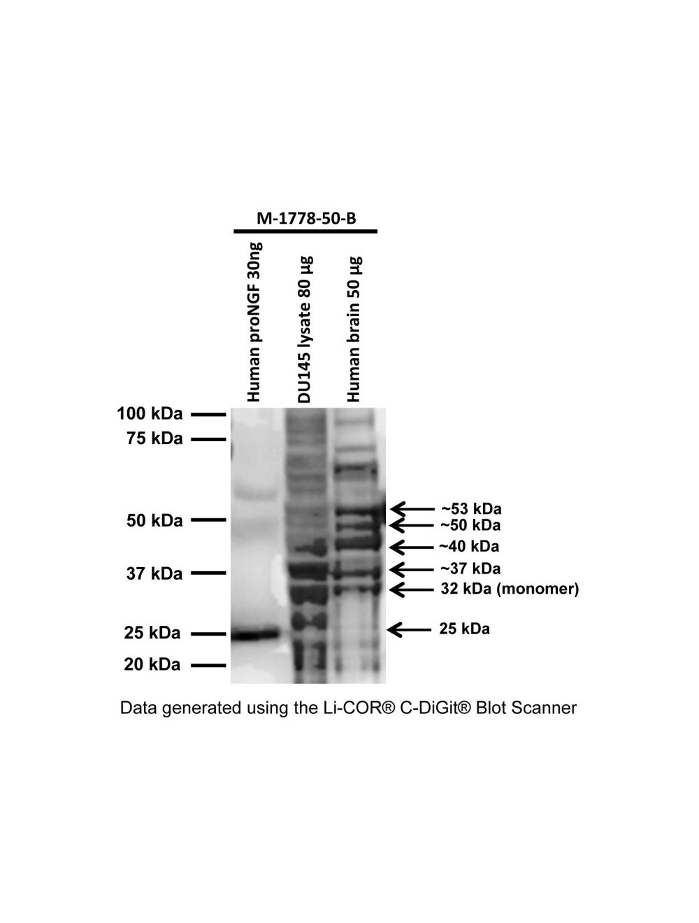

Pro-Nerve growth factor (proNGF), Mouse Monoclonal Antibody (Biotin)
- Product Name Pro-Nerve growth factor (proNGF), Mouse Monoclonal Antibody (Biotin)
-
Product Description
Mouse anti-Pro-Nerve growth factor (proNGF) Monoclonal Antibody (Biotin), suitable for WB.
- Alternative Names Pro-brain nerve growth factor; proNGF; NGF
- Application(s) WB
- Antibody Host Mouse
- Antibody Type Monoclonal
- Specificity Human Species cross-reactivity not tested.
- Species Reactivity Human
- Immunogen Description A synthetic peptide (C-HTIPQAHWTKLQ, aa: 30-41) of human proNGF protein has been used as the immunogen. The sequence is located on the pro-domain of the proNGF full-length protein and is 80% homologous to mouse and rat proNGF.
- Conjugate Biotin
- Purity Description Antibody was purified from cell culture supernatant by Protein G chromatography, biotinylated and buffer-exchanged into PBS, pH 7.4 buffer
- Regulatory Status For research use only.
Product Info
-
Product Description
Mouse anti-Pro-Nerve growth factor (proNGF) Monoclonal Antibody (Biotin), suitable for WB.
-
Related Products
Pro-nerve growth factor (proNGF), Mouse Monoclonal Antibody
- Application(s) WB
-
Application Details
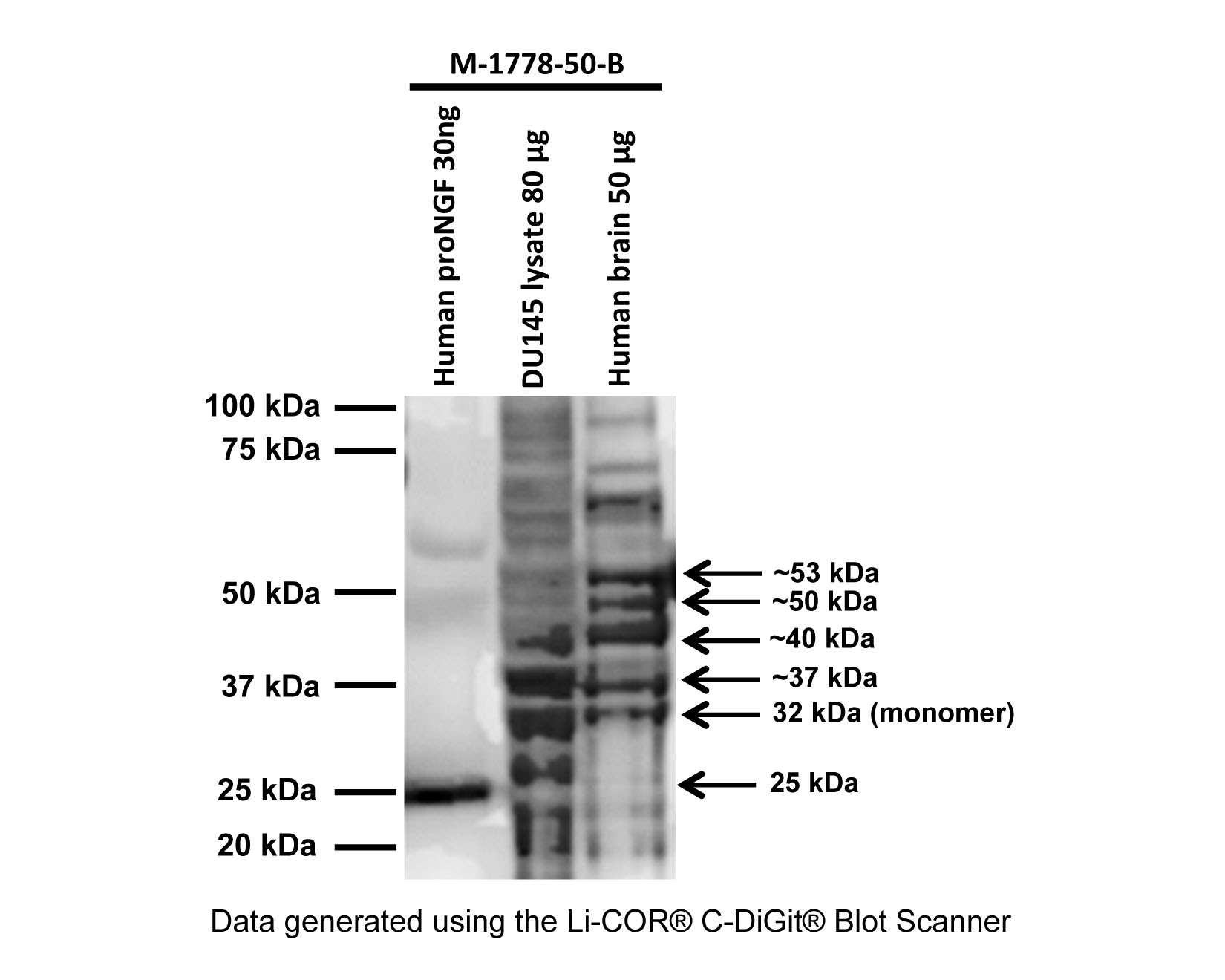
The biotinylated proNGF antibody has been tested by Western Blotting (0.1-0.5 µg/mL) and is also expected to work in applications validated for the unlabelled antibody M-1738-100 at same or higher dilutions: Flow Cytometry and Immunofluorescence.
Other applications not yet tested. Biosensis recommends optimal dilutions/concentrations should be determined by the end user. - Target Pro-Nerve growth factor (proNGF)
- Specificity Human Species cross-reactivity not tested.
- Target Host Species Human
- Species Reactivity Human
- Antibody Host Mouse
- Antibody Type Monoclonal
- Antibody Isotype IgG2, lambda
- Clone Name BS312
- Conjugate Biotin
- Immunogen Description A synthetic peptide (C-HTIPQAHWTKLQ, aa: 30-41) of human proNGF protein has been used as the immunogen. The sequence is located on the pro-domain of the proNGF full-length protein and is 80% homologous to mouse and rat proNGF.
- Purity Description Antibody was purified from cell culture supernatant by Protein G chromatography, biotinylated and buffer-exchanged into PBS, pH 7.4 buffer
- Format Lyophilized from a solution containing PBS buffer pH 7.4 with 3% trehalose, without preservatives.
- Reconstitution Instructions Spin vial briefly before opening. Reconstitute in 50 µL of sterile-filtered, ultrapure water to give a concentration of 1 mg/mL. Centrifuge to remove any insoluble material. Final buffer contains no preservative.
- Storage Instructions After reconstitution divide into aliquots and store at -20°C for a higher stability. Antibody contains no preservatives. Store at 2-8°C with an appropriate antibacterial agent. Use sterile methods. Highest purity Glycerol (1:1) may be added for an additional stability when stored at refrigerated or freezing temperatures. Avoid repetitive freeze/thaw cycles.
- Batch Number Please see item label.
- Expiration Date 12 months after date of receipt (unopened vial).
- Alternative Names Pro-brain nerve growth factor; proNGF; NGF
- Uniprot Number P01138
- Uniprot Number/Name P01138 (NGF_HUMAN)
-
Scientific Background
Nerve growth factor (NGF) is synthesized as a precursor (proNGF) which may be released and have physiological functions to cause cell death. It binds neurotrophin receptor p75 and sortilin and may also be important for the development of nervous system. proNGF is synthesized in target tissues and glia, transported retrogradely and may be released.
Biosensis now offers biotinylated proNGF antibody allowing more flexibility in experimental design by using the biotin-avidin/streptavidin detection method. The ability of biotinylated proNGF antibody to detect proNGF has been validated by WB. - Shipping Temperature 25°C (ambient)
- UNSPSC CODE 41116161
- Regulatory Status For research use only.
Specifications
-
Specific References
Makoudjou M et al. (2024) ProNGF processing in adult rat tissues and bioactivity of NGF pro domain peptides FEBSPRESS Application: Rat Brain Tissue