1800 605-5127
 +61 (0)8 8352 7711
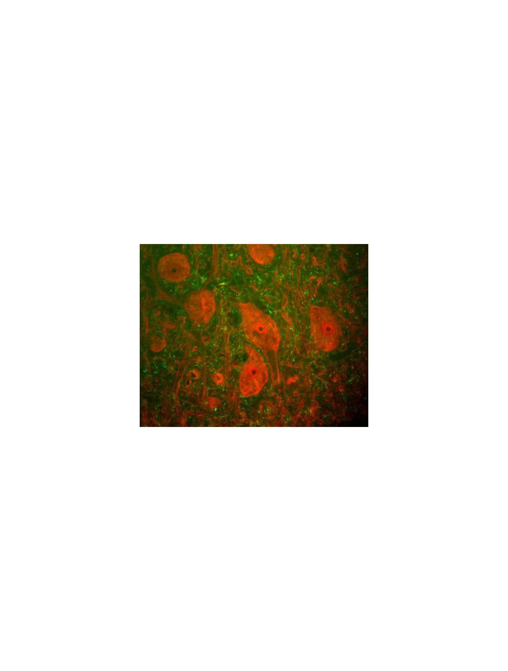

Ubiquitin carboxyl-terminal hydrolase isozyme L1 (UCHL1), Mouse Monoclonal Antibody
- Product Name Ubiquitin carboxyl-terminal hydrolase isozyme L1 (UCHL1), Mouse Monoclonal Antibody
- Product Description Mouse anti-Ubiquitin carboxyl-terminal hydrolase isozyme L1 (UCHL1) Monoclonal Antibody (Unconjugated), suitable for WB, IHC-Frozen, ICC.
- Alternative Names Ubiquitin carboxyl-terminal hydrolase isozyme L1; UCH-L1; Neuron cytoplasmic protein 9.5; PGP 9.5; PGP9.5; Ubiquitin thioesterase L1; UCHL1;
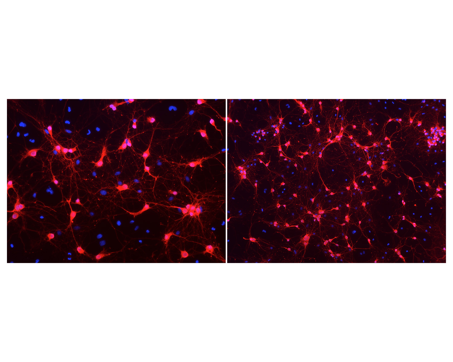
- Application(s) ICC, IHC-Frozen, WB
- Antibody Host Mouse
- Antibody Type Monoclonal
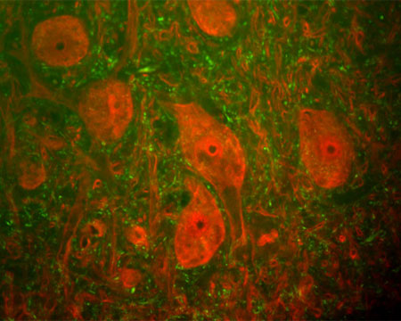
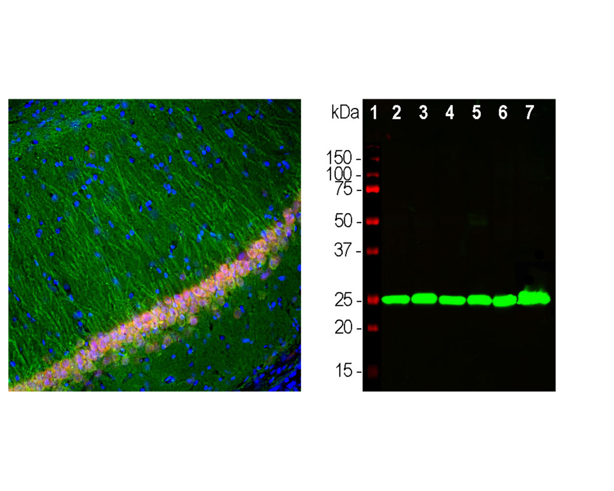
- Specificity The specificity of this antibody has been confirmed by WB. This antibody detects ~24 kDa UCHL1 enzyme. Suitable control tissue is rat spinal cord, brain, SHSY-5Y or HEK293 cell extract. Hu, Rat, Bov
- Species Reactivity Bovine, Human, Mouse, Pig, Rat
- Immunogen Description Recombinant full length human Ubiquitin C Terminal Hydrolase 1 (UCHL1) purified from E. coli.
- Conjugate Unconjugated
- Purity Description Protein G purified
- Regulatory Status For research use only.
Product Info
- Product Description Mouse anti-Ubiquitin carboxyl-terminal hydrolase isozyme L1 (UCHL1) Monoclonal Antibody (Unconjugated), suitable for WB, IHC-Frozen, ICC.
- Application(s) ICC, IHC-Frozen, WB
- Application Details Western Blotting (WB), Immunocytochemistry (ICC) and Immunohistochemistry (IHC). A dilution of 1:10,000 - 1:20,000 is recommended for WB. A dilution of 1:1,000 - 1:5,000 is recommended for IC and IH. Biosensis recommends optimal dilutions/concentrations should be determined by the end user.
- Target Ubiquitin carboxyl-terminal hydrolase isozyme L1 (UCHL1)
- Specificity The specificity of this antibody has been confirmed by WB. This antibody detects ~24 kDa UCHL1 enzyme. Suitable control tissue is rat spinal cord, brain, SHSY-5Y or HEK293 cell extract. Hu, Rat, Bov
- Target Host Species Human
- Species Reactivity Bovine, Human, Mouse, Pig, Rat
- Antibody Host Mouse
- Antibody Type Monoclonal
- Antibody Isotype IgG1
- Clone Name BH7
- Conjugate Unconjugated
- Immunogen Description Recombinant full length human Ubiquitin C Terminal Hydrolase 1 (UCHL1) purified from E. coli.
- Purity Description Protein G purified
- Format Lyophilized from PBS buffer pH 7.2-7.6 with 0.1% trehalose, and sodium azide
- Reconstitution Instructions Spin vial briefly before opening. Reconstitute with 100 µL sterile-filtered, ultrapure water to achieve a 1 mg/mL concentration. Centrifuge to remove any insoluble material.
- Storage Instructions After reconstitution of lyophilized antibody, aliquot and store at -20°C for a higher stability. Avoid freeze-thaw cycles.
- Batch Number Please see item label.
- Expiration Date 12 months after date of receipt (unopened vial).
- Alternative Names Ubiquitin carboxyl-terminal hydrolase isozyme L1; UCH-L1; Neuron cytoplasmic protein 9.5; PGP 9.5; PGP9.5; Ubiquitin thioesterase L1; UCHL1;
- Uniprot Number P09936
- Uniprot Number/Name P09936 (UCHL1_HUMAN)
- Scientific Background This enzyme is a thiol protease that recognizes and hydrolyzes a peptide bond at the C-terminal glycine of ubiquitin. The enzyme also binds to free monoubiquitin and may prevent its degradation in lysosomes (ref: SWISSPROT).
- Shipping Temperature 25°C (ambient)
- UNSPSC CODE 41116161
- Regulatory Status For research use only.