SpecificityThe antibody reacts with multiple closely spaced bands covering the region of the blot from 48 kDa to 67 kDa, with an additional band at 100 kDa. It has also been used successfully for immunocytochemistry. Expected to react with horse, cow, pig, chicken, rat and mouse.
Species ReactivityBovine (Predicted), Chicken (Predicted), Horse (Predicted), Human, Mouse (Predicted), Pig (Predicted), Rat (Predicted)
Immunogen DescriptionRecombinant full length version of the shortest human tau isoform purified from E. coli.
Chicken anti-Microtubule-associated protein tau (MAPT) Polyclonal Antibody (Unconjugated), suitable for WB, ICC.
Application(s)ICC, WB
Application DetailsWestern Blotting (WB) and Immunocytochemistry (ICC). A dilution of 1:5,000 - 1:10,000 is recommended for WB. A dilution of 1:500-1,000 is recommended for IC. Biosensis recommends optimal dilutions/concentrations should be determined by the end user.
TargetMicrotubule-associated protein tau (MAPT)
SpecificityThe antibody reacts with multiple closely spaced bands covering the region of the blot from 48 kDa to 67 kDa, with an additional band at 100 kDa. It has also been used successfully for immunocytochemistry. Expected to react with horse, cow, pig, chicken, rat and mouse.
Target Host SpeciesHuman
Species ReactivityBovine (Predicted), Chicken (Predicted), Horse (Predicted), Human, Mouse (Predicted), Pig (Predicted), Rat (Predicted)
Antibody HostChicken
Antibody TypePolyclonal
Antibody IsotypeIgY
ConjugateUnconjugated
Immunogen DescriptionRecombinant full length version of the shortest human tau isoform purified from E. coli.
Purity DescriptionIgY
FormatLyophilized IgY preparation, with sodium azide.
Reconstitution InstructionsSpin vial briefly before opening. Reconstitute with 100 µL sterile-filtered, ultrapure water. Centrifuge to remove any insoluble material.
Storage InstructionsStore lyophilized, unopened vial at 2-8°C or lower. After reconstitution, prepare aliquots and store at -20°C to -80°C for a higher stability. Avoid freeze-thaw cycles.
Batch NumberPlease see item label.
Expiration Date12 months after date of receipt (unopened vial).
Scientific BackgroundFUNCTION: Promotes microtubule assembly and stability, and might be involved in the establishment and maintenance of neuronal polarity. The C-terminus binds axonal microtubules while the N-terminus binds neural plasma membrane components, suggesting that tau functions as a linker protein between both. Axonal polarity is predetermined by tau localization (in the neuronal cell) in the domain of the cell body defined by the centrosome. The short isoforms allow plasticity of the cytoskeleton whereas the longer isoforms may preferentially play a role in its stabilization. SUBCELLULAR LOCATION: Cytoplasm; cytosol. Cell membrane. Mostly found in the axons of neurons, in the cytosol and in association with plasma membrane components. ALTERNATIVE PRODUCTS: 8 named isoforms produced by alternative splicing. Additional isoforms seem to exist. Isoforms differ from each other by the presence or absence of up to 5 of the 15 exons. One of these optional exons contains the additional tau/MAP repeat. TISSUE SPECIFICITY: Expressed in neurons. Isoform PNS-tau is expressed in the peripheral nervous system while the others are expressed in the central nervous system. DEVELOPMENTAL STAGE: Four-repeat (type II) tau is expressed in an adult-specific manner and is not found in fetal brain, whereas three-repeat (type I) tau is found in both adult and fetal brain. DOMAIN: The tau/MAP repeat binds to tubulin. In Alzheimer disease, the neuronal cytoskeleton in the brain is progressively disrupted and replaced by tangles of paired helical filaments and straight filaments, mainly composed of hyperphosphorylated forms of Microtubule-associated protein Tau. Defects in Microtubule-associated protein Tau are a cause of frontotemporal dementia and parkinsonism linked to chromosome 17, as well as a number of other neurodegenerative diseases.
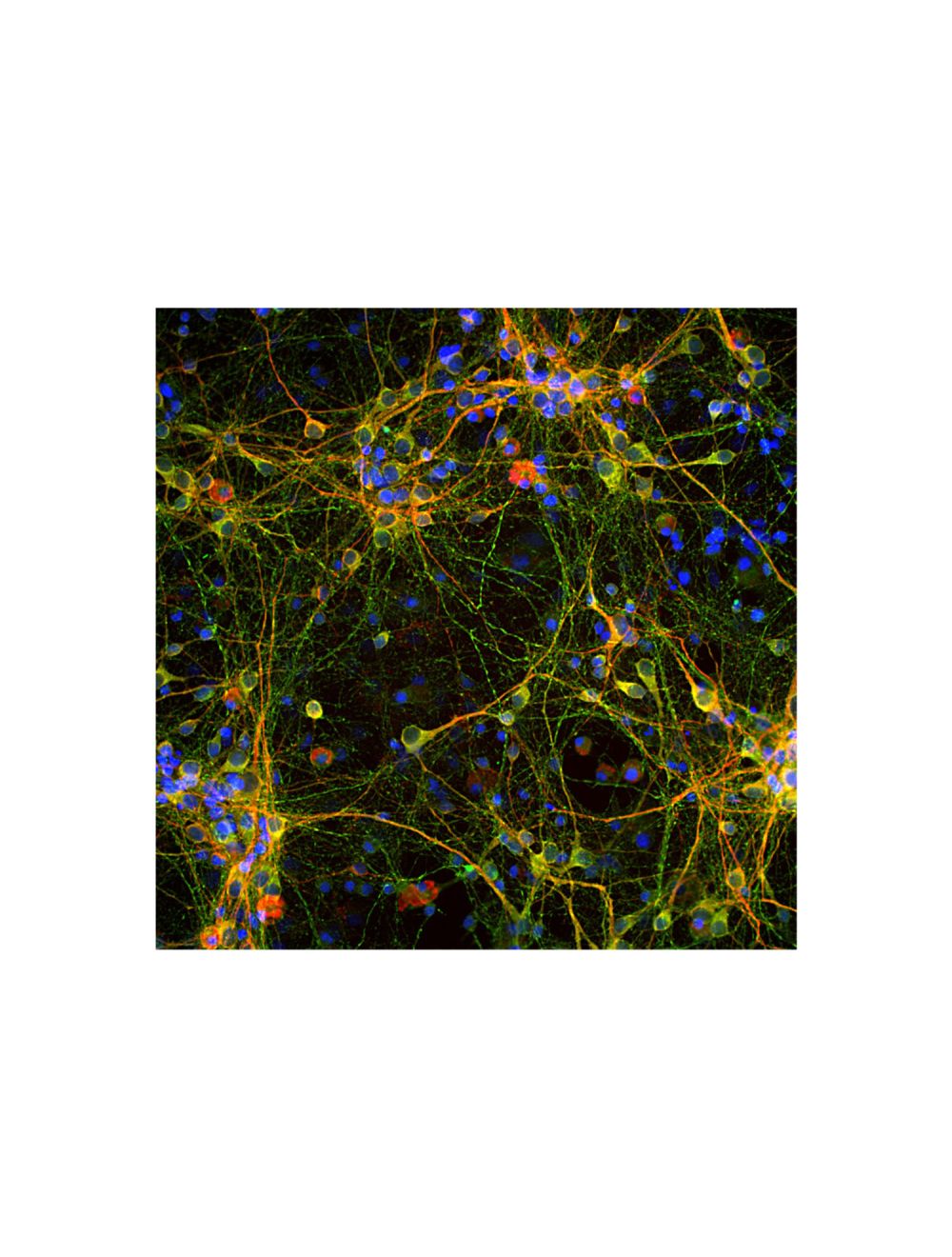
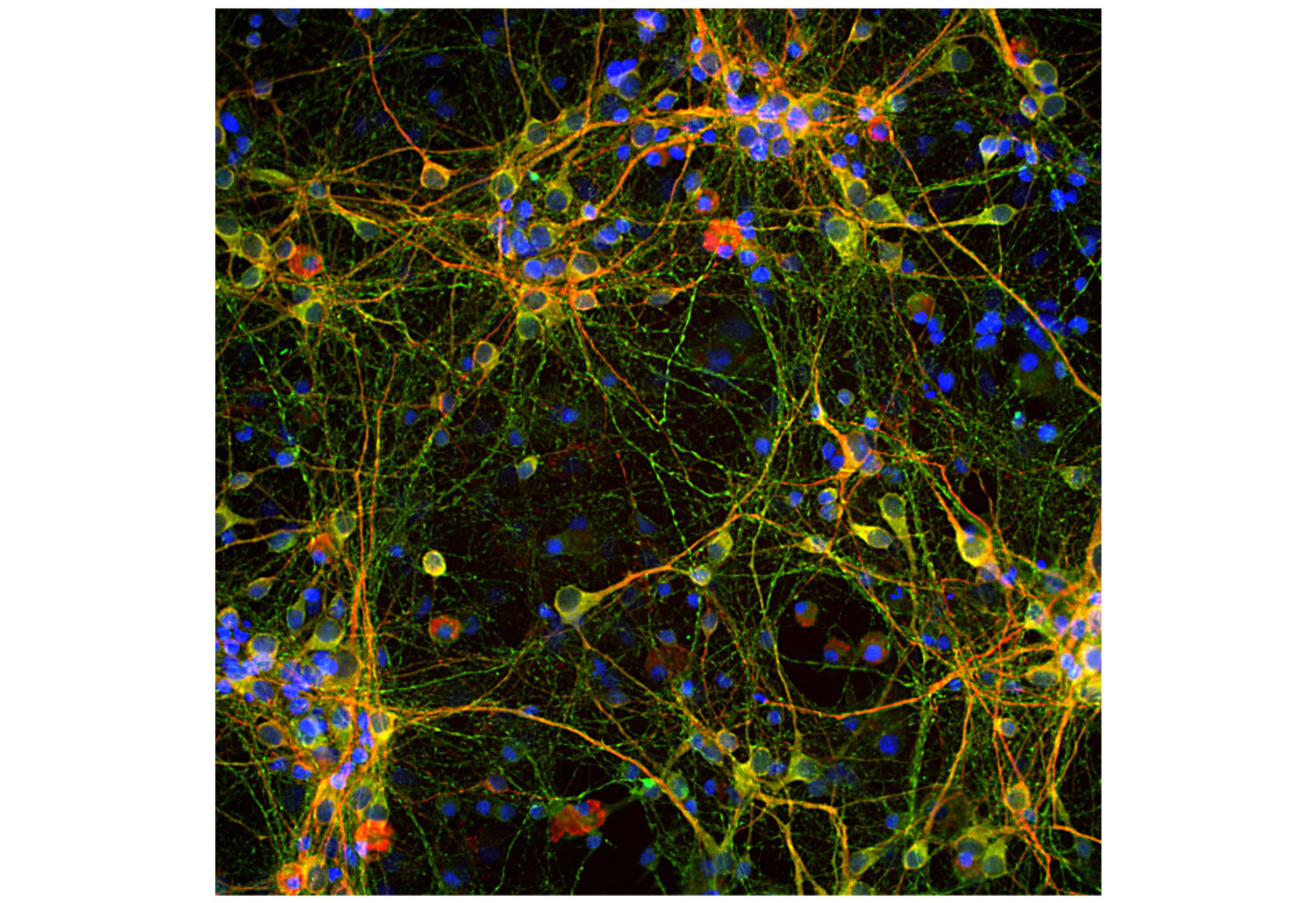
Immunofluorescence analysis of tau protein expression in cortical neuron-glial culture from E20 rat. The chicken anti-tau antibody (green, 1:2,000) stains perikarya, dendrites and axons of neurons, while a anti-MAP2 antibody labels only dendrites and perikarya (red). Perikarya and dendrites appear orange-yellow, since they contain both MAP2 and tau proteins. Blue: DAPI nuclear stain.
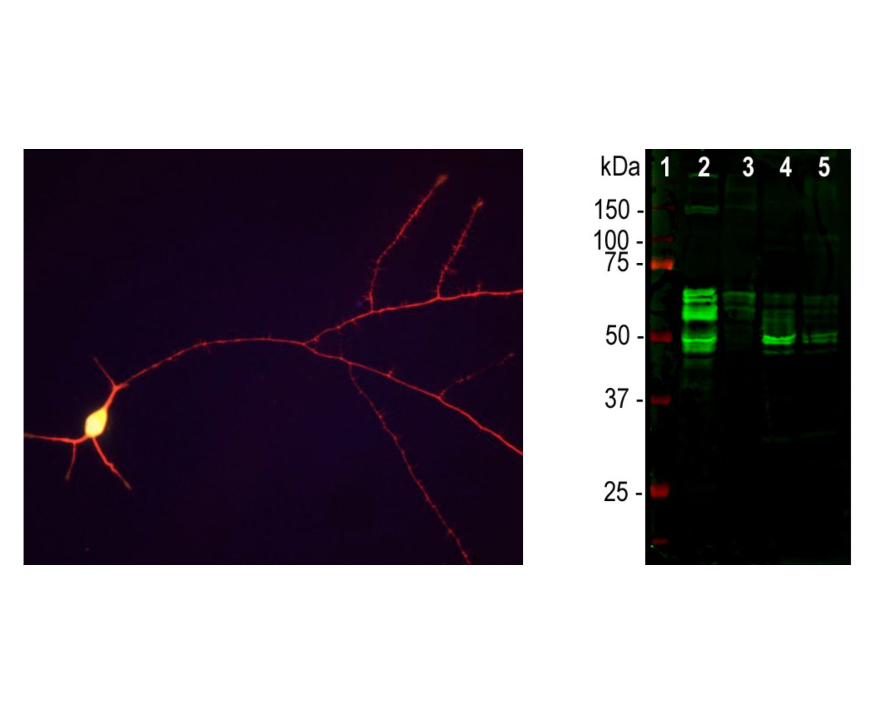
Left: Immunofluorescence staining of E18 hippocampal neurons with chicken anti-Tau antibody C-1691-100 (red). Ubiquitin C-terminal Hydrolase 1 (UCHL1), an abundant cytoplasmic protein of neurons which is concentrated in the perikarya, was double-labelled with M-1407-100. Since the perikarya contain both UCHL1 and Tau, the red and green signals superimpose resulting in yellow color. Neuronal processes, which contain relatively much more tau, appear red. Cell nuclei were stained with DAPI (blue). Right: Western blot analysis of tau protein expression in tissue (antibody dilution: 1:10,000). [1] protein standard, [2] rat brain, [3] rat spinal cord, [4] mouse brain, [5] mouse spinal cord. Tau protein is expressed as multiple isoforms of different molecular weight, and so appears as multiple closely spaced bands in the region of the blot in the range from 48 kDa to 67 kDa.
Specific ReferencesChen J et al. (2023) APP mediates tau uptake and its overexpression leads to the exacerbated tau pathology Cell Mol Life Sci. 80(5):123 Application: IHC/IF Species: Mouse
General ReferencesSkene JH, Willard M. Changes in axonally transported proteins during axon regeneration in toad retinal ganglion cells. J. Cell Biol. 89:86-95 (1981).
 1800 605-5127
 +61 (0)8 8352 7711