Product NameMicrotubule-associated protein tau (MAPT), Mouse Monoclonal Antibody
Product DescriptiongoogleMouse anti-Microtubule-associated protein tau (MAPT) Monoclonal Antibody (Unconjugated), suitable for WB, ICC.
Alternative NamesNeurofibrillary tangle protein; Paired helical filament-tau; PHF-tau; MAPT; MTBT1; TAU
Application(s)ICC, WB
Antibody HostMouse
Antibody TypeMonoclonal
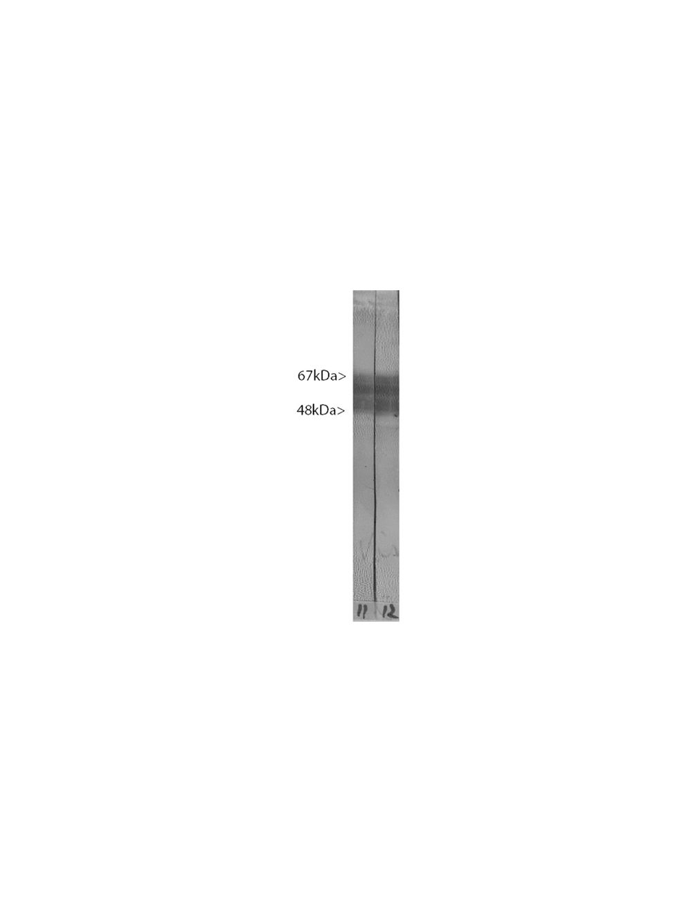
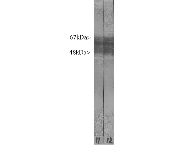
SpecificityThe antibody reacts with multiple closely spaced bands covering the region of the blot from 48 kDa to 67 kDa. It has also been used successfully for immunocytochemistry.
Species ReactivityHuman, Mouse, Other Mammals (Predicted), Rat
Immunogen DescriptionRecombinant full length version of the shortest human tau isoform purified from E. coli.
Product DescriptionMouse anti-Microtubule-associated protein tau (MAPT) Monoclonal Antibody (Unconjugated), suitable for WB, ICC.
Application(s)ICC, WB
Application DetailsWestern Blotting (WB) and Immunocytochemistry (ICC). A dilution of 1:5,000 - 1:10,000 is recommended for WB. A dilution of 1:500-1,000 is recommended for ICC. Biosensis recommends optimal dilutions/concentrations should be determined by the end user.
TargetMicrotubule-associated protein tau (MAPT)
SpecificityThe antibody reacts with multiple closely spaced bands covering the region of the blot from 48 kDa to 67 kDa. It has also been used successfully for immunocytochemistry.
Target Host SpeciesHuman
Species ReactivityHuman, Mouse, Other Mammals (Predicted), Rat
Antibody HostMouse
Antibody TypeMonoclonal
Antibody IsotypeIgG1
Clone Name2E9
ConjugateUnconjugated
Immunogen DescriptionRecombinant full length version of the shortest human tau isoform purified from E. coli.
Purity DescriptionAffinity Purified
FormatLyophilized from PBS buffer pH 7.2-7.6 with 0.1% trehalose, and sodium azide
Reconstitution InstructionsSpin vial briefly before opening. Reconstitute with 100 µL sterile-filtered, ultrapure water to achieve a 1 mg/mL concentration. Centrifuge to remove any insoluble material.
Storage InstructionsMaintain lyophilized material at 2-8°C. After reconstitution of lyophilized antibody, aliquot and store at -20°C for a higher stability. Avoid freeze-thaw cycles.
Batch NumberPlease see item label.
Expiration Date12 months after date of receipt (unopened vial).
Alternative NamesNeurofibrillary tangle protein; Paired helical filament-tau; PHF-tau; MAPT; MTBT1; TAU
Scientific BackgroundFUNCTION: Promotes microtubule assembly and stability, and might be involved in the establishment and maintenance of neuronal polarity. The C-terminus binds axonal microtubules while the N-terminus binds neural plasma membrane components, suggesting that tau functions as a linker protein between both. Axonal polarity is predetermined by tau localization (in the neuronal cell) in the domain of the cell body defined by the centrosome. The short isoforms allow plasticity of the cytoskeleton whereas the longer isoforms may preferentially play a role in its stabilization. SUBCELLULAR LOCATION: Cytoplasm; cytosol. Cell membrane. Mostly found in the axons of neurons, in the cytosol and in association with plasma membrane components. ALTERNATIVE PRODUCTS: 8 named isoforms produced by alternative splicing. Additional isoforms seem to exist. Isoforms differ from each other by the presence or absence of up to 5 of the 15 exons. One of these optional exons contains the additional tau/MAP repeat. TISSUE SPECIFICITY: Expressed in neurons. Isoform PNS-tau is expressed in the peripheral nervous system while the others are expressed in the central nervous system. DEVELOPMENTAL STAGE: Four-repeat (type II) tau is expressed in an adult-specific manner and is not found in fetal brain, whereas three-repeat (type I) tau is found in both adult and fetal brain. DOMAIN: The tau/MAP repeat binds to tubulin. In Alzheimer disease, the neuronal cytoskeleton in the brain is progressively disrupted and replaced by tangles of paired helical filaments and straight filaments, mainly composed of hyperphosphorylated forms of Microtubule-associated protein Tau. Defects in Microtubule-associated protein Tau are a cause of frontotemporal dementia and parkinsonism linked to chromosome 17, as well as a number of other neurodegenerative diseases.
Crude rat brain extract. Tau protein is expressed as up to 9 different isoforms of different molecular weight and so appears as multiple closely spaced bands covering the region of the blot from 48 kDa to 67 kDa.
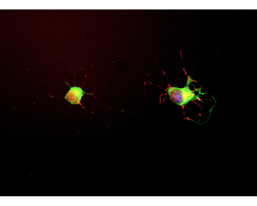
E18 hippocampal neurons were grown for seven days. The neurons were fixed and immunostained with M-1703-100 (red channel). The cells were also stained in green with C-1377-50, our chicken antibody to alpha Internexin and DNA (blue). M-1703-100 stains the neuronal perikarya and process strongly, and does not stain non neuronal cells in these cultures. The alpha internexin antibody stains intermediate or 10nm filament bundles in the cytoplasm of these cells.
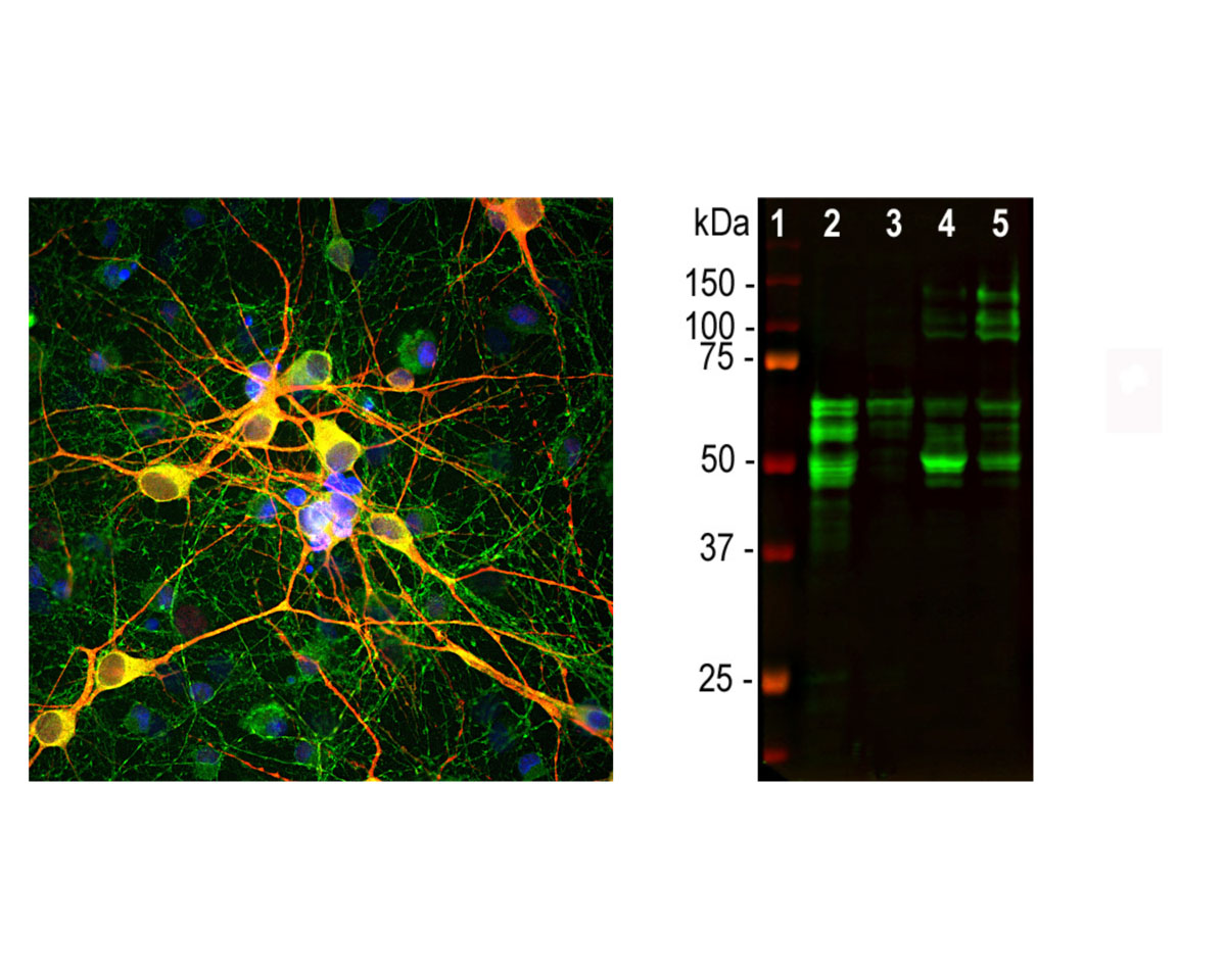
Left: Analysis of MAP-tau expression (green) in cortical neuron-glial culture from E20 rat by Immunocytochemistry. Mouse antibody to MAP-tau was used at 1:1,000 dilution. Co-staining was performed with chicken antibody to MAP2 (C-1382-50, red, 1:5,000). Blue: DAPI nuclear stain. The MAP-tau antibody stains perikarya, dendrites and axons of neurons, while the MAP2 antibody labels only dendrites and perikarya. As a result, perikarya and dendrites appear orange-yellow, since they contain both proteins. Right: Western blot analysis of MAP-tau expression in tissue lysates using mouse antibody to MAP-tau (green, 1:2,000). [1] protein standard, [2] rat brain, [3] rat spinal cord, [4] mouse brain, [5] mouse spinal cord. Tau protein is expressed as up to 9 different isoforms of different molecular weights, therefore, it appears as multiple closely spaced bands in the range from 48 kDa to 67 kDa in the CNS (lanes 2-4). In the PNS, additional higher molecular weight bands are observed (lane 5).
General ReferencesSkene JH, Willard M. Changes in axonally transported proteins during axon regeneration in toad retinal ganglion cells. J. Cell Biol. 89:86-95 (1981).
 1800 605-5127
 +61 (0)8 8352 7711