SpecificityThe specificity of this antibody has been confirmed by WB. This antibody detects ~8.5 kDa Ubiquitin. Hu, Bov, Chk, Drosophila, and C. elegans
Species ReactivityBovine, C. elegans, Chicken, Drosophila, Human
Immunogen DescriptionRaised against purified ubiquitin conjugated with glutaraldehyde to keyhole limpet hemocyanin.
Application DetailsWestern Blotting (WB), Immunohistochemistry - paraffin embedded tissue (IH-P) and ELISA. Suggested dilution for WB is 1:500-1,000. This antibody can be used on mildly fixed histological sections of human brain for studies of Alzheimer's disease. This antibody also works on paraffin embedded material. It also recognises other ubiquinated inclusion bodies such as Lewy bodies of Parkinson's disease and the Pick bodies in Pick's disease in formalin fixed tissues. Suggested dilution for IH is 1:500. Biosensis recommends optimal dilutions/concentrations should be determined by the end user.
TargetPolyubiqutin-B
SpecificityThe specificity of this antibody has been confirmed by WB. This antibody detects ~8.5 kDa Ubiquitin. Hu, Bov, Chk, Drosophila, and C. elegans
Target Host SpeciesHuman
Species ReactivityBovine, C. elegans, Chicken, Drosophila, Human
Antibody HostMouse
Antibody TypeMonoclonal
Antibody IsotypeIgG1
Clone NameUbi-1
ConjugateUnconjugated
Immunogen DescriptionRaised against purified ubiquitin conjugated with glutaraldehyde to keyhole limpet hemocyanin.
Purity DescriptionProtein G purified
FormatLyophilized from PBS buffer pH 7.2-7.6 with 0.1% trehalose, and sodium azide
Reconstitution InstructionsSpin vial briefly before opening. Reconstitute with 100 µL sterile-filtered, ultrapure water to achieve a 1 mg/mL concentration. Centrifuge to remove any insoluble material.
Storage InstructionsAfter reconstitution of lyophilized antibody, aliquot and store at -20°C for a higher stability. Avoid freeze-thaw cycles.
Batch NumberPlease see item label.
Expiration Date12 months after date of receipt (unopened vial).
Alternative NamesRPS27A; UBA52; UBB; UBC; Polyubiquitin-B; Polyubiquitin-C;
Scientific BackgroundUbiquitin is a highly conserved 76 amino acid protein with an estimated molecular weight of 8.56 kDa which has a central role in regulated protein degradation. It is a protein modifier which can be covalently attached to target lysines either as a monomer or as a lysine-linked polymer. Several types of polymeric chains can be formed depending on the lysine used for the assembly. Attachment to proteins as a polymer leads to their degradation by the 26S proteosome; a complex, multicatalytic cytosolic and nuclear protease. Attachment to proteins as a monomer or as an alternatively linked polymer does not lead to proteasomal degradation and may be required for numerous functions, including maintenance of chromatic structure, regulation of gene expression, stress response, ribosome biogenesis and DNA repair. Ubiquitin is synthesized as a polyubiquitin precursor with exact head to tail repeats, the number of repeats of which differ between species and strains. In some species there is a final amino-acid after the last repeat, here in bovine a Cys. Some ubiquitin genes contain a single copy of ubiquitin fused to a ribosomal protein (either L40 or S27a).
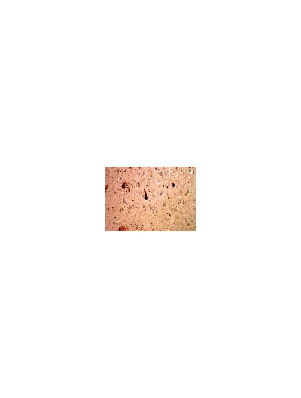
Mouse monoclonal antibody to Ubiquitin [Ubi-1] M-1404-100 staining of cerebral cortex of an Alzheimer patient. Neurofibrillary tangles and dystrophic neurites associated with senile plaques stain strongly with this antibody. In the center is a typical neurofibrillary tangle containing neuron.
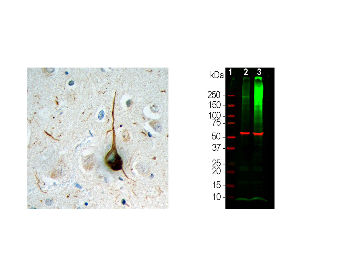
Left: Analysis of ubiquitin expression (brown) in FFPE section of human cerebral cortex of an Alzheimer patient by Immunohistochemistry. Signal was generated with DAB. Counterstain: haemotoxylin (blue). A typical flame-shaped tangle is seen in a pyramidal neuron in the center and is surrounded by dystrophic neurites, also strongly ubiquitin-positive. Both are commonly seen in cortical and hippocampal Alzheimer brain sections and are typical for this disease, but are rare or absent in healthy brain. Right: Western blot analysis of HEK293 cell lysates for ubiquitin expression (green, 1:1,000). [1] protein standard, [2] cells maintained in normal medium, [3] cells treated with 10 uM of proteasome inhibitor lactacystin (Lc) for 16 hours. The smear detected above the 200 kDa standard represents accumulation of ubiquitinated proteins in proteasome inhibitor-Lc treated cells. The prominent band at 8 kDa corresponds to monoubiquitin. The same blot was probed with an antibody to HSP60 as loading control.
 1800 605-5127
 +61 (0)8 8352 7711