Product NameVisinin-like protein 1 (VLP-1), Mouse Monoclonal Antibody
Product DescriptiongoogleMouse anti-Visinin-like protein 1 (VLP-1) Monoclonal Antibody (Unconjugated), suitable for WB, ICC, IHC-Frozen.
Alternative NamesHippocalcin-like protein 3, HLP3, HPCAL3, HUVISL1, VLP-1, VILIP and VILIP-1
Application(s)ICC, IHC-Frozen, WB
Antibody HostMouse
Antibody TypeMonoclonal
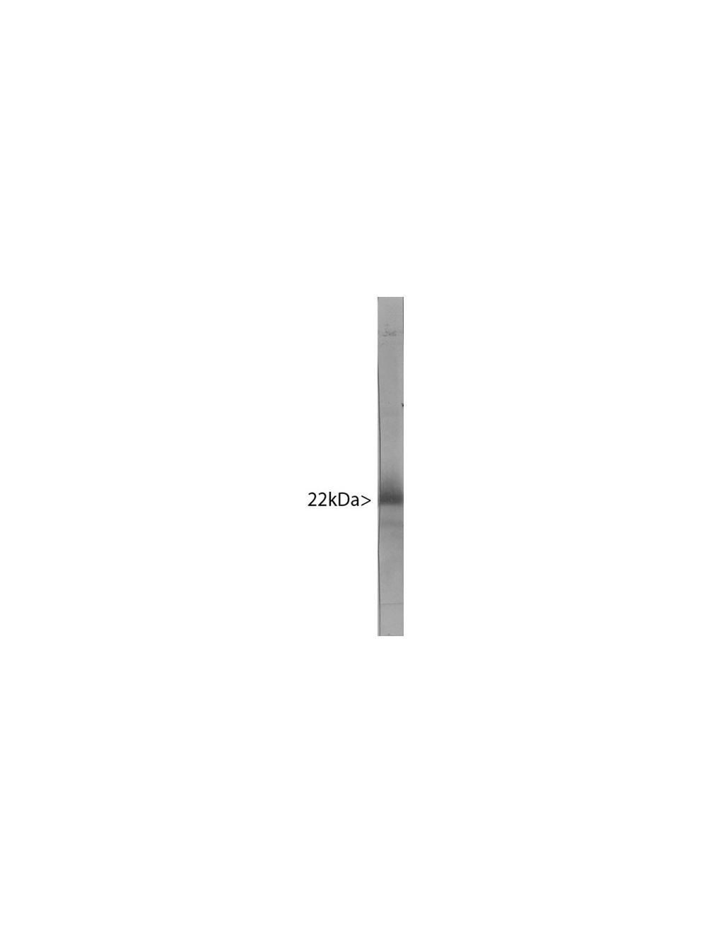
SpecificityThe antibody reacts with a 22 kDa band by Western blot on bovine cerebellum lysate. It has also been used successfully for immunocytochemistry.
Species ReactivityBovine, Human, Mouse, Other Mammals (Predicted), Rat
Immunogen DescriptionRecombinant Visinin-like protein 1 expressed and purified from E. coli.
Product DescriptionMouse anti-Visinin-like protein 1 (VLP-1) Monoclonal Antibody (Unconjugated), suitable for WB, ICC, IHC-Frozen.
Application(s)ICC, IHC-Frozen, WB
Application DetailsWestern Blotting (WB), Immunocytochemistry (ICC) and Immunohistochemistry (IHC). A dilution of 1:500 - 1:1,000 is recommended for WB. A dilution of 1:500-1:1,000 is recommended for IHC and ICC. Biosensis recommends optimal dilutions/concentrations should be determined by the end user.
TargetVisinin-like protein 1 (VLP-1)
SpecificityThe antibody reacts with a 22 kDa band by Western blot on bovine cerebellum lysate. It has also been used successfully for immunocytochemistry.
Target Host SpeciesHuman
Species ReactivityBovine, Human, Mouse, Other Mammals (Predicted), Rat
Antibody HostMouse
Antibody TypeMonoclonal
Antibody IsotypeIgG1
Clone Name2D11
ConjugateUnconjugated
Immunogen DescriptionRecombinant Visinin-like protein 1 expressed and purified from E. coli.
Purity DescriptionProtein G purified
FormatLyophilized from PBS buffer pH 7.2-7.6 with 0.1% trehalose, and sodium azide
Reconstitution InstructionsSpin vial briefly before opening. Reconstitute with 100 µL sterile-filtered, ultrapure water to achieve a 1 mg/mL concentration. Centrifuge to remove any insoluble material.
Storage InstructionsAfter reconstitution of lyophilized antibody, aliquot and store at -20°C for a higher stability. Avoid freeze-thaw cycles.
Batch NumberPlease see item label.
Expiration Date12 months after date of receipt (unopened vial).
Alternative NamesHippocalcin-like protein 3, HLP3, HPCAL3, HUVISL1, VLP-1, VILIP and VILIP-1
Scientific BackgroundVisinin (sometimes known as hippocalcin-like protein 3, HLP3, HPCAL3, HUVISL1, VLP-1, VILIP and VILIP-1) was originally isolated biochemically from chicken retina as a major protein of about 24 kDa on SDS-PAGE (1). Following cloning and sequencing of visinin, several visinin like proteins were discovered by homology screening (2, 3). One of these, Visinin-like protein 1 is a small Calcium binding protein which is very abundant in the nervous system and is found only in neurons, though different neurons have different levels of expression (4, 5). It is particularly concentrated in cerebellar Purkinje cells, and tends to be most abundant in perikarya and dendrites. The protein belongs to the large superfamLy of calmodulin and paravalbumin type proteins which function by binding Calcium ions. Calcium binding alters the confomation of these proteins and allow them to interact with other binding partners, the properties of which they may alter. Visinin-like protein 1 has four "EF hand" domains, which are negatively charged helix-turn-helix peptides which are responsible for Calcium binding. Visinin-like protein 1 is 191 amino acids in size and has a molecular weight on SDS-PAGE of 22 kDa. The protein has recently been suggested to be a useful biomarker of Alzheimer's disease and traumatic brain injury (6, 7, 8).
Western blot of bovine cerebellum homogenate stained with M-1657-100.
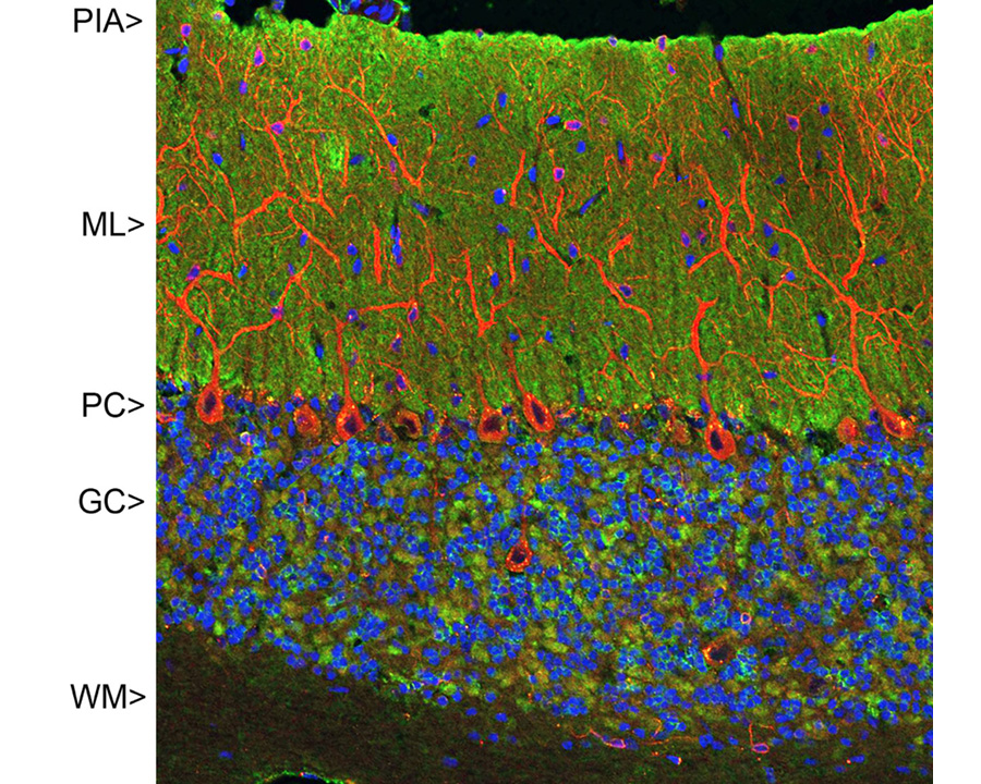
Confocal image of adult rat cerebellar cortex stained with M-1657-100 (green), our chicken polyclonal antibody to MAP2 (Catalog Number C-1382-50, red) and DNA (blue). The M-1657-100 antibody reveals synapses in the molecular layer (ML) strongly. Synaptic regions are also seen in the granule cell layer (GC). The perikarya of Purkinje cells (PC) are revealed with MAP2 antibody (4). Little staining is seen in the white matter (WM).
Left: VILIP-1 staining of rat cerebellum section by Immunohistochemistry. Section was stained with mouse antibody to VILIP-1 (red, 1:500) and rabbit antibody to GFAP (R-1374-50, green, 1:5,000). Blue: DAPI nuclear stain.IHC Method: Following transcardial perfusion of rat with 4% paraformaldehyde, brain was post-fixed for 24 hours, cut to 45 um, and free-floating sections were stained. The VILIP-1 antibody reveals protein expressed in granule cell membranes and in synapses in the white matter and molecular layers of the cerebellum. The GFAP antibody stains the processes of Bergmann glia and astroglia. Right: Western blot analysis of VILIP-1 expression (green, 1:1,000) in tissue lysates. [1] protein standard, [2] rat brain [3] rat cerebellum, and [4] mouse brain. The band at around 20 kDa corresponds to VILIP-1 protein.
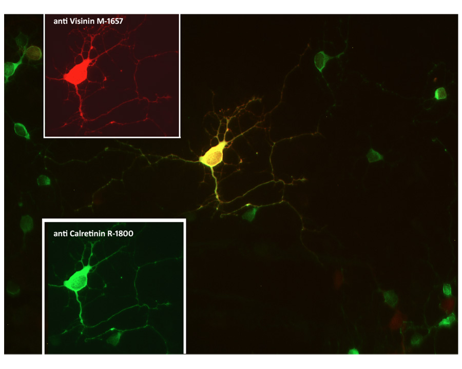
Co-localization of calretinin (green) and visinin (red) in retinal neuron visualized by Immunocytochemistry. Rat retinal primary cells (QBM cat# R-Ret-508) were cultured for 7 days. Image courtesy of QBM Cell Science.
Staining of calretinin (red) and VILIP1 (green) positive neurons by Immunocytochemistry in rat retina culture. Rat retinal primary cells (QBM cat# R-Ret-508) were cultured for 7 days. Image courtesy of QBM Cell Science.
General ReferencesHatakenaka S, Kuo CH, Miki N. Analysis of a distinctive protein in chick retina during development. Brain Res. 312:155-63 (1983). Kuno T, Kajimoto Y, Hashimoto T, Mukai H, Shirai Y, Saheki S, Tanaka C. cDNA cloning of a neural visinin-like Ca(2+)-binding protein. Biochem Biophys Res Commun. 184:1219-25 (1992). Polymeropoulos MH, Ide S, Soares MB, Lennon GG. Sequence characterization and genetic mapping of the human VSNL1 gene, a homologue of the rat visinin-like peptide RNVP1. Genomics. 29:273-5 (1995). Bernstein HG, Baumann B, Danos P, Diekmann S, Bogerts B, Gundelfinger ED, Braunewell KH. Regional and cellular distribution of neural visinin-like protein immunoreactivities (VILIP-1 and VILIP-3) in human brain. J Neurocytol. 28:655-62 (1999). Paterlini M, Revilla V, Grant AL, Wisden W Expression of the neuronal calcium sensor protein family in the rat brain. .Neuroscience. 99:205-16 (2000) Laterza OF, Modur VR, Crimmins DL, Olander JV, Landt Y, Lee JM, Ladenson JH. Identification of novel brain biomarkers. Clin. Chem. 9:1713-21 (2006) Lee JM, Blennow K, Andreasen N, Laterza O, Modur V, Olander J, Gao F, Ohlendorf M, Ladenson JH. The brain injury biomarker VLP-1 is increased in the cerebrospinal fluid of Alzheimer disease patients. Clin. Chem. 10:1617-23 (2008). Tarawneh R, D'Angelo G, Macy E, Xiong C, Carter D, Cairns NJ, Fagan AM, Head D, Mintun MA, Ladenson JH, Lee JM, Morris JC, Holtzman DM. Visinin-like protein-1: diagnostic and prognostic biomarker in Alzheimer disease. Ann Neurol. 70:274-85 (2011) doi: 10.1002/ana.22448.
 1800 605-5127
 +61 (0)8 8352 7711