Product NameNeurofilament Heavy, phosphorylated and non-phosphorylated (pNF-H), Rabbit Polyclonal Antibody
Product DescriptiongoogleRabbit anti-Neurofilament Heavy, phosphorylated and non-phosphorylated, (pNF-H) Polyclonal Antibody (Unconjugated), suitable for WB and Immunostaining.
Alternative NamesNF-H; NFH; NF-200; NF200; NF-H; NEFH; N52; Neurofilament heavy polypeptide; Neurofilament triplet H protein; 200 kDa neurofilament protein; KIAA0845
Application(s)IF, ICC, WB
Antibody HostRabbit
Antibody TypePolyclonal
SpecificitySpecies cross-reactivity includes human and rat. This antibody reacts with with phosphorylated NF-H at approx 200 kDa and non-phosphorylated NF-H at 160 kDa. Predicted to react with other mammals due to sequence homology.
Species ReactivityHuman, Other Mammals (Predicted), Rat
Immunogen DescriptionThis antibody has been made against a rat NF-H construct containing most of the tandem KSP repeats expressed in and purified from E.coli.
Application DetailsWestern blot (WB) and Immunocytochemistry (ICC) / Immunofluorescence (IF). A dilution of 1:5,000-10,000 is recommended for WB. A dilution of 1:500-1,000 is recommended for ICC/IF. This antibody stains dendritic and perikaryal neurofilaments particularly well. Biosensis recommends optimal dilutions/concentrations should be determined by the end user.
TargetNeurofilament heavy polypeptide , phosphorylated and non-phosphorylated (pNF-H)
SpecificitySpecies cross-reactivity includes human and rat. This antibody reacts with with phosphorylated NF-H at approx 200 kDa and non-phosphorylated NF-H at 160 kDa. Predicted to react with other mammals due to sequence homology.
Target Host SpeciesRat
Species ReactivityHuman, Other Mammals (Predicted), Rat
Antibody HostRabbit
Antibody TypePolyclonal
Antibody IsotypeMixed
ConjugateUnconjugated
Immunogen DescriptionThis antibody has been made against a rat NF-H construct containing most of the tandem KSP repeats expressed in and purified from E.coli.
Purity DescriptionWhole serum
FormatLyophilized with sodium azide.
Reconstitution InstructionsSpin vial briefly before opening. Reconstitute with 50 µL sterile-filtered, ultrapure water. Centrifuge to remove any insoluble material.
Storage InstructionsStore lyophilized antibody at 2-8°C After reconstitution of lyophilized antibody, aliquot and store at -20°C for a higher stability. Avoid freeze-thaw cycles. Store at 4°C for up to one month for short term storage and frequent use.
Batch NumberPlease see item label.
Expiration Date12 months after date of receipt (unopened vial).
Alternative NamesNF-H; NFH; NF-200; NF200; NF-H; NEFH; N52; Neurofilament heavy polypeptide; Neurofilament triplet H protein; 200 kDa neurofilament protein; KIAA0845
Scientific BackgroundNeurofilaments are the 10nm or intermediate filament proteins found specifically in neurons, and are composed predominantly of three major proteins called NF-L, NF-M and NF-H, though other filament proteins may be included also. The major function of neurofilaments is likely to control the diameter of large axons. NF-L is the neurofilament light or low molecular weight polypeptide and runs on SDS-PAGE gels at 68-70kDa with some variability across species. Antibodies to NF-L are useful for identifying neuronal cells and their processes in cell culture and sectioned material. NF-L antibody can also be useful for the visualization of neurofilament rich accumulations seen in many neurological diseases, such as Lou Gehrig’s disease (ALS), giant axon neuropathy, Charcot-Marie Tooth disease and others. (Ref: uniprot.org)
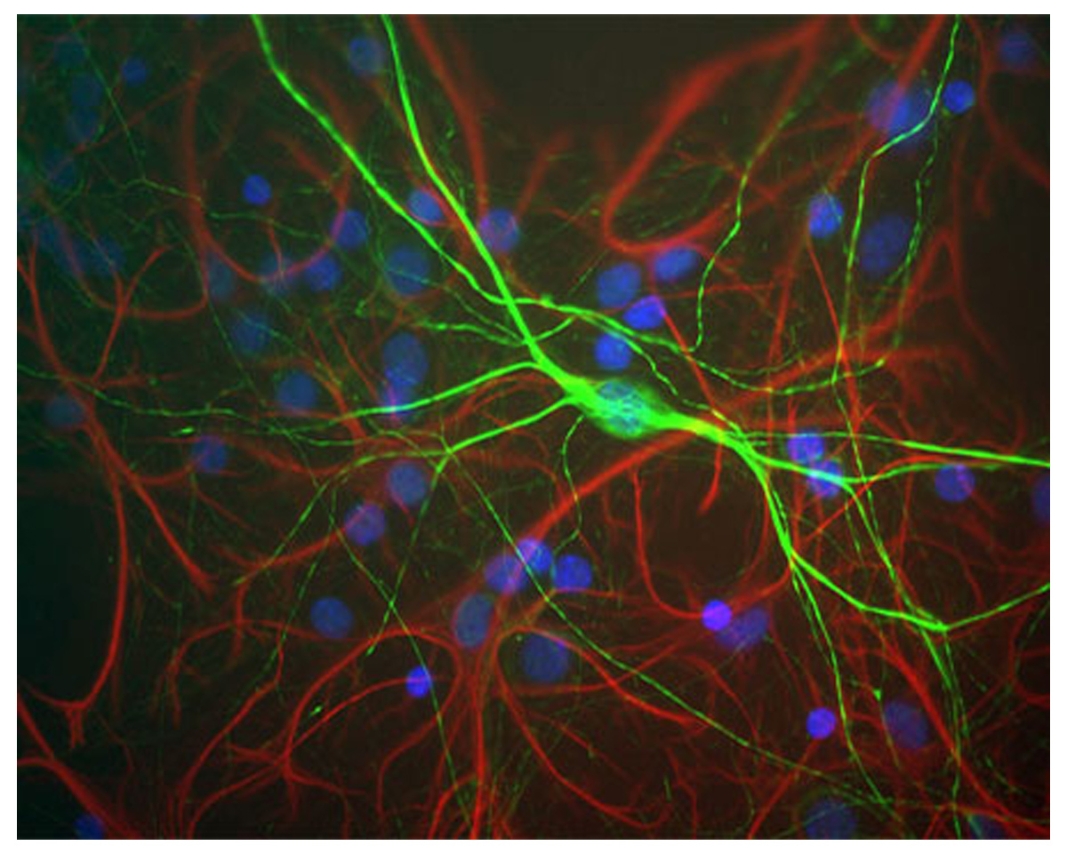
Image of mixed neuron and glial cultures stained by Immunofluorescence and Immunocytochemistry. The cultures were stained with R-1389-50, Neurofilament heavy polypeptide, phosphorylated and non-phosphorylated (pNF-H), Rabbit pAb (green), and co-stained with product C-1373-50, Glial Fibrillary Acidic Protein (GFAP), Chicken pAb, (red). The neurofilament NF-H antibody binds to phosphorylated axonal forms of NF-H and non-phosphorylated dendritic and perikaryal forms.
Image of membrane TrkC expression (green) in human HeLa cells by Immunofluorescence and Immunocytochemistry. Method: Fixed (4% formaldehyde) and blocked (10% normal horse serum) HeLa cells were incubated with product M-1837-100, Tyrosine Kinase Receptor C (TrkC), Clone BS337, Mouse mAb, (green, 2 µg/mL) for 1 hour. Cells were then permeabilized, blocked (10% horse serum, 0.1% Triton X-100) and incubated with product R-1389-50 (red, 1:500) for 1 hour. Primary antibody binding was visualized with secondary donkey anti-mouse-CF488A and donkey anti-rabbit-CF568 antibodies (2 µg/mL, 1 hour incubation). Cell nuclei were stained with DAPI (blue). Negative control staining was performed under identical conditions with isotype control mouse antibody and rabbit whole serum from an non-immunized animal. Confocal imaging settings were the same for antibody and control staining. Magnification: 60x. Specific staining is observed for TrkC in the cell membrane, while the NF-H antibody reveals intracellular filaments.
Image of of membrane TrkC expression (green) in human HeLa cells by 3D confocal imaging . TrkC-IR is concentrated in the elongated protrusion away from the cell body, while intracellular NF-H expression (red) is observed throughout the cell. The blue is DAPI staining of nuclear DNA
 1800 605-5127
 +61 (0)8 8352 7711