Product NameNeurofilament heavy polypeptide, phosphorylated, (pNF-H), Rabbit Polyclonal Antibody
Product DescriptiongoogleRabbit anti-Neurofilament heavy polypeptide, phosphorylated, (pNF-H), Polyclonal Antibody (Unconjugated), suitable for WB and Immunostaining.
Alternative NamesNF-H; NFH; NF-200; NF200; NF-H; NEFH; N52; Neurofilament heavy polypeptide; Neurofilament triplet H protein; 200 kDa neurofilament protein; KIAA0845
Application(s)IF, ICC, IHC, WB
Antibody HostRabbit
Antibody TypePolyclonal
SpecificitySpecies cross-reactivity includes human, rat, mouse, cow, pig and horse. This antibody reacts with phosphorylated NF-H and is seen as a band at approx 200 kDa in WB. Predicted to react with other mammals due to sequence homology.
Species ReactivityBovine, Horse, Human, Mouse, Other Mammals (Predicted), Pig, Rat
Immunogen DescriptionThis antibody has been made against biochemically isolated NF-H purified from bovine spinal cord.
Product DescriptionRabbit anti-Neurofilament heavy polypeptide, phosphorylated, (pNF-H), Polyclonal Antibody (Unconjugated), suitable for WB and Immunostaining.
Application DetailsWestern blot (WB), Immunocytochemistry (ICC) / Immunofluorescence (IF), and Immunocytochemistry (IHC). A dilution of 1:10,000-25,000 is recommended for WB. A dilution of 1:1,000-1:5,000 is recommended for ICC/IF andIHC. Biosensis recommends optimal dilutions/concentrations should be determined by the end user.
TargetNeurofilament heavy polypeptide, phosphorylated (pNF-H)
SpecificitySpecies cross-reactivity includes human, rat, mouse, cow, pig and horse. This antibody reacts with phosphorylated NF-H and is seen as a band at approx 200 kDa in WB. Predicted to react with other mammals due to sequence homology.
Target Host SpeciesBovine
Species ReactivityBovine, Horse, Human, Mouse, Other Mammals (Predicted), Pig, Rat
Antibody HostRabbit
Antibody TypePolyclonal
Antibody IsotypeMixed
ConjugateUnconjugated
Immunogen DescriptionThis antibody has been made against biochemically isolated NF-H purified from bovine spinal cord.
Purity DescriptionWhole serum
FormatLyophilized with sodium azide.
Reconstitution InstructionsSpin vial briefly before opening. Reconstitute with 50 µL sterile-filtered, ultrapure water. Centrifuge to remove any insoluble material.
Storage InstructionsStore lyophilized antibody at 2-8°C After reconstitution of lyophilized antibody, aliquot and store at -20°C for a higher stability. Avoid freeze-thaw cycles. Store at 4°C for up to one month for short term storage and frequent use.
Batch NumberPlease see item label.
Expiration Date12 months after date of receipt (unopened vial).
Alternative NamesNF-H; NFH; NF-200; NF200; NF-H; NEFH; N52; Neurofilament heavy polypeptide; Neurofilament triplet H protein; 200 kDa neurofilament protein; KIAA0845
Scientific BackgroundNeurofilaments are the 10nm or intermediate filament proteins found specifically in neurons, and are composed predominantly of three major proteins called NF-L, NF-M and NF-H, though other filament proteins may be included also. The major function of neurofilaments is likely to control the diameter of large axons. NF-L is the neurofilament light or low molecular weight polypeptide and runs on SDS-PAGE gels at 68-70kDa with some variability across species. Antibodies to NF-L are useful for identifying neuronal cells and their processes in cell culture and sectioned material. NF-L antibody can also be useful for the visualization of neurofilament rich accumulations seen in many neurological diseases, such as Lou Gehrig’s disease (ALS), giant axon neuropathy, Charcot-Marie Tooth disease and others. (Ref: uniprot.org)
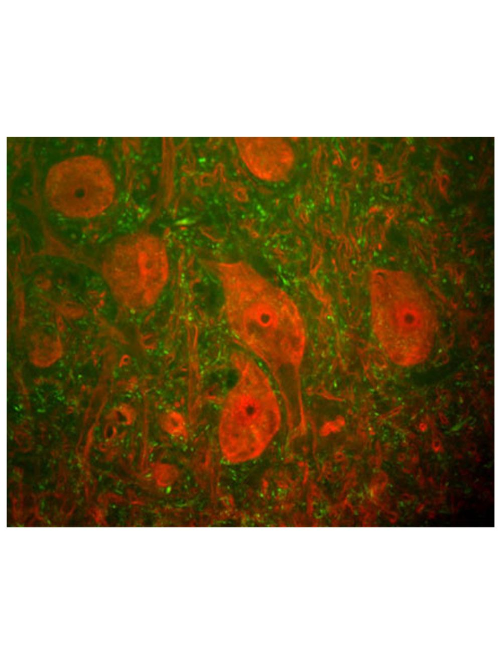
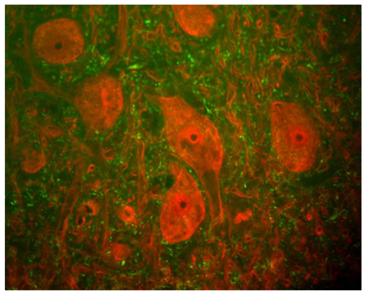
Image of a rat spinal cord section by Immunofluorescence and Immunohistochemistry. The section was stained with R-1388-50, Neurofilament heavy polypeptide, phosphorylated (pNF-H), Rabbit pAb, (green) and co-stained with product M-1407-100, Ubiquitin carboxyl-terminal hydrolase isozyme L1 (UCHL1), Mouse mAb, (red). The neurofilament NF-H antibody binds primarily to phosphorylated axonal forms of NF-H, and so stains axons coursing between the large UCHL1 positive neurons. These large cells are a-motorneurons and UCHL1 protein is a major component of the perikarya and dendrites of these cells.
Image of a mouse hippocampus section by Immunofluorescence and Immunohistochemistry. The section was stained with R-1388-50, Neurofilament heavy polypeptide, phosphorylated, (pNF-H), Rabbit pAb, (red, 1:2,000) and co-stained with product M-1384-50, Myelin basic protein (MBP), Clone 7G7, Mouse mAb,(green, 1:5,000). The blue is DAPI staining of nuclear DNA. Method: Following transcardial perfusion with 4% paraformaldehyde, brain was post-fixed for 24 hours, cut to 45 um, and free-floating sections were stained. The NF-H antibody labels a network of axons of different neurons, while the MBP antibody stains myelin sheath around these axons.
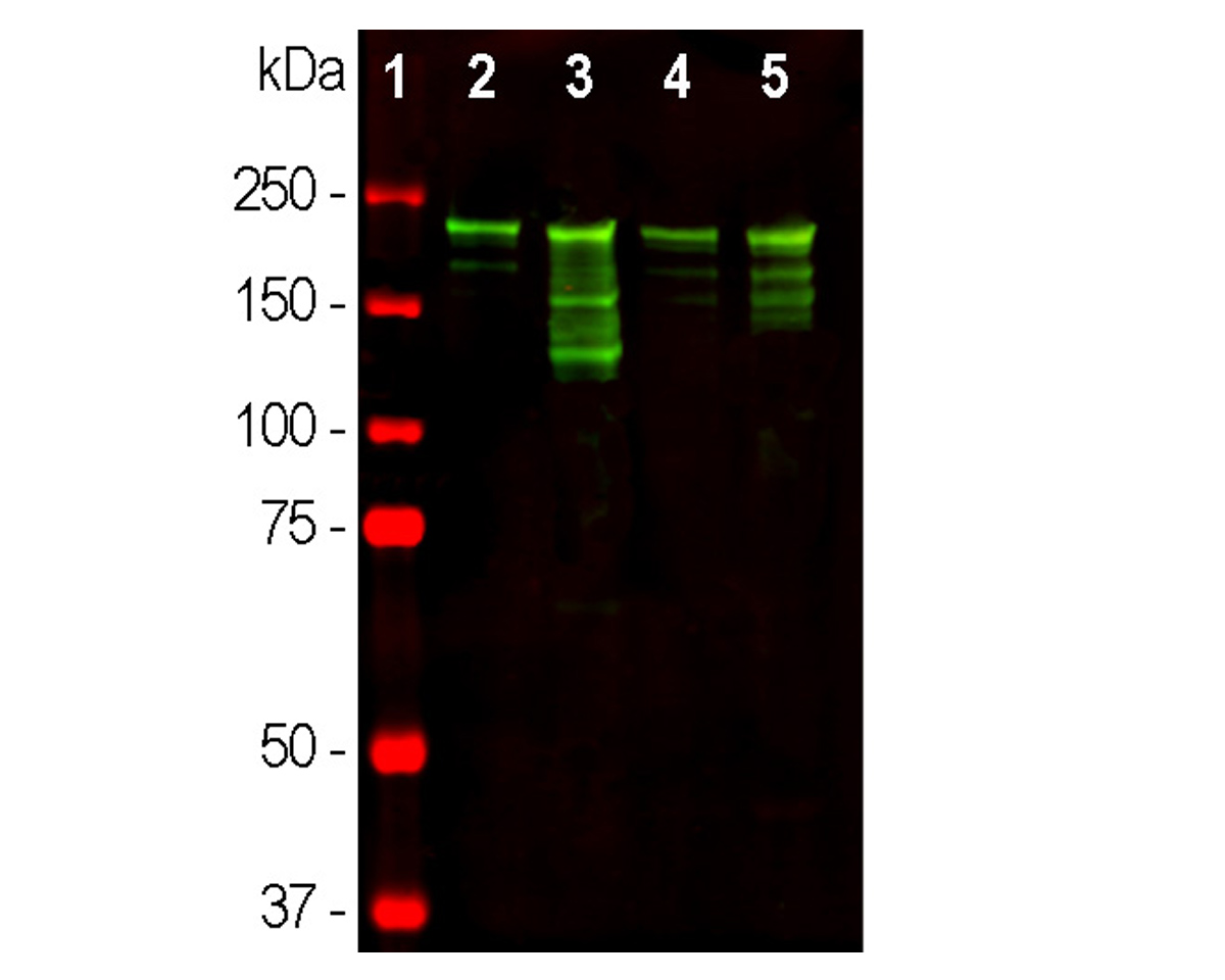
Analysis by western blot of pNF-H expression in tissue lysates with R-1388-50, Neurofilament heavy polypeptide, phosphorylated, (pNF-H), Rabbit pAb (green 1:10,000) with a strong band at spprox 220 kDa corresponding to the phosphorylated axonal form of the NF-H subunit. Smaller proteolytic fragments of NF-H are also detected with this antibody. Lane 1: protein standard; Lane 2: rat brain; Lane 3: rat spinal cord; Lane 4: mouse brain; Lane 5: mouse spinal cord lysate.
 1800 605-5127
 +61 (0)8 8352 7711