1800 605-5127
 +61 (0)8 8352 7711
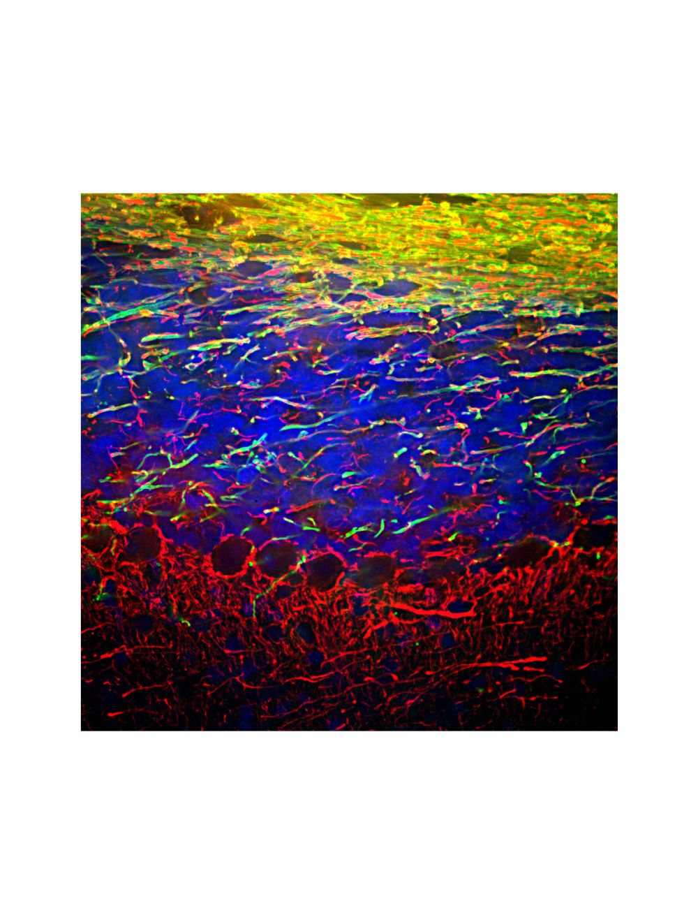

Neurofilament light polypeptide (NF-L), Rabbit Polyclonal Antibody
- Product Name Neurofilament light polypeptide (NF-L), Rabbit Polyclonal Antibody
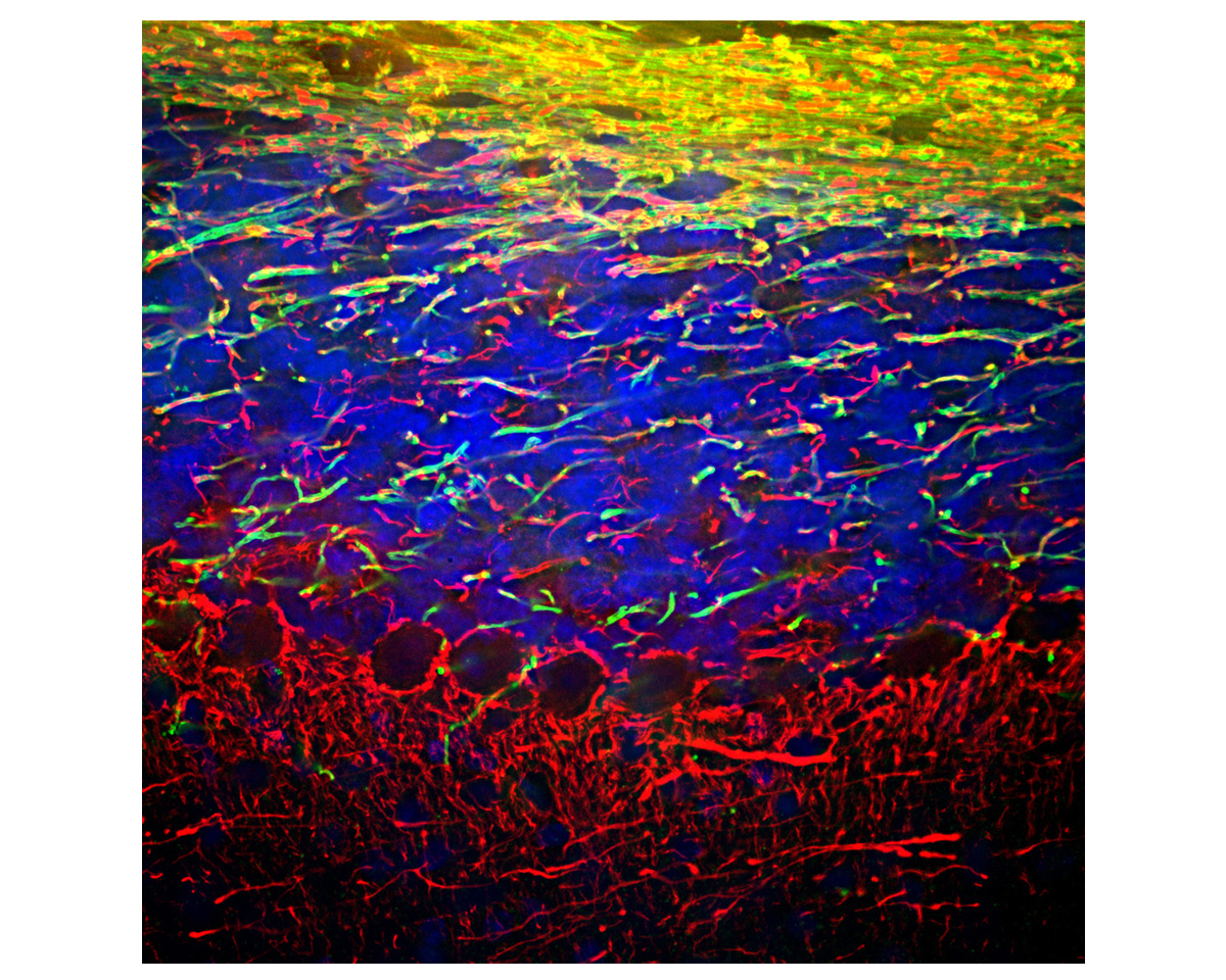
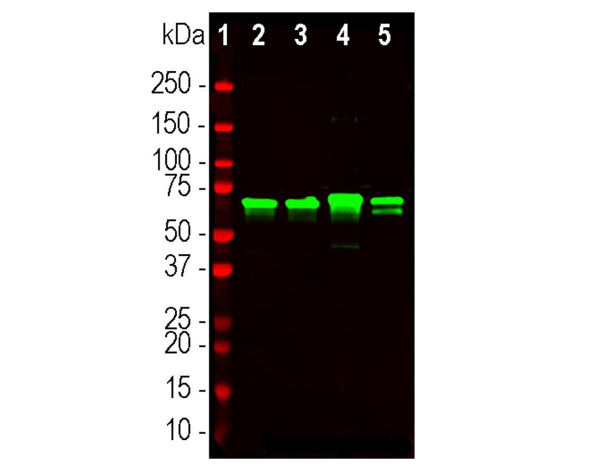
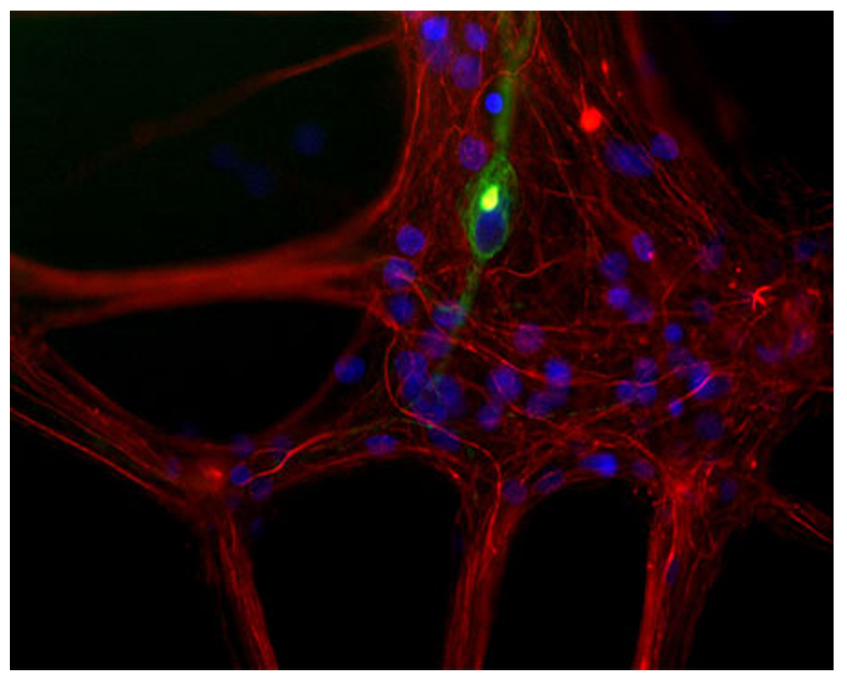
- Product Description Rabbit anti-Neurofilament light polypeptide (NF-L), Polyclonal Antibody (Unconjugated), suitable for WB and Immunostaining.
- Alternative Names NF-L; NF68; NEFL; Neurofilament light polypeptide; NFL; neurofilament light chain; Neurofilament triplet L protein; NFL
- Application(s) IF, ICC, IHC, WB
- Antibody Host Rabbit
- Antibody Type Polyclonal
- Specificity Species cross-reactivity includes human, rat, mouse, cow, and pig.
- Species Reactivity Bovine, Human, Mouse, Pig, Rat
- Immunogen Description The antibody has been made against a preparation of recombinant full length human NF-L. The antibody binds NF-L from a variety of species including human, rat and mouse.
- Conjugate Unconjugated
- Purity Description Whole serum
- Regulatory Status For research use only.
Product Info
- Product Description Rabbit anti-Neurofilament light polypeptide (NF-L), Polyclonal Antibody (Unconjugated), suitable for WB and Immunostaining.
-
Related Products
Neurofilament light polypeptide (NF-L), Chicken Polyclonal Antibody
Neurofilament light polypeptide (NF-L), DG-Sensor™, Chicken Polyclonal Antibody
Neurofilament light polypeptide (NF-L), Mouse Monoclonal Antibody (DA2)
Neurofilament light polypeptide (NF-L), Mouse Monoclonal Antibody (7D1)
Neurofilament light polypeptide (NF-L), Mouse Monoclonal Antibody (6H112)
Neurofilament light polypeptide (NF-L), DG-Sensor™, Mouse Monoclonal Antibody (6H63)
Neurofilament light polypeptide (NF-L), DG-Sensor™, Mouse Monoclonal Antibody (1D44)
Neurofilament light polypeptide (NF-L), Mouse Monoclonal Antibody (1B11)
Neurofilament light polypeptide, C-terminus, (NF-L-Ct), Rabbit Polyclonal Antibody
Neurofilament light polypeptide (NF-L), DG-Sensor™, Rabbit Polyclonal Antibody
- Application(s) IF, ICC, IHC, WB
- Application Details Western blot (WB), Immunocytochemistry (ICC) / Immunofluorescence (IF), Immunohistochemistry (IHC). A dilution of 1:10,000 – 1:15,000 is recommended for WB. A dilution of 1:2,000 - 1:5,000 is recommended for ICC/IF and a dilution of 1:5,000 is recommended for IHC. Biosensis recommends optimal dilutions/concentrations should be determined by the end user.
- Target Neurofilament light polypeptide (NF-L)
- Specificity Species cross-reactivity includes human, rat, mouse, cow, and pig.
- Target Host Species Human
- Species Reactivity Bovine, Human, Mouse, Pig, Rat
- Antibody Host Rabbit
- Antibody Type Polyclonal
- Antibody Isotype Mixed
- Conjugate Unconjugated
- Immunogen Description The antibody has been made against a preparation of recombinant full length human NF-L. The antibody binds NF-L from a variety of species including human, rat and mouse.
- Purity Description Whole serum
- Format Lyophilized with sodium azide.
- Reconstitution Instructions Spin vial briefly before opening. Reconstitute with 50 µL sterile-filtered, ultrapure water. Centrifuge to remove any insoluble material.
- Storage Instructions Store lyophilized antibody at 2-8°C After reconstitution of lyophilized antibody, aliquot and store at -20°C for a higher stability. Avoid freeze-thaw cycles. Store at 4°C for up to one month for short term storage and frequent use.
- Batch Number Please see item label.
- Expiration Date 12 months after date of receipt (unopened vial).
- Alternative Names NF-L; NF68; NEFL; Neurofilament light polypeptide; NFL; neurofilament light chain; Neurofilament triplet L protein; NFL
- Uniprot Number P07196
- Uniprot Number/Name P07196 (NFL_HUMAN)
- Scientific Background Neurofilaments are composed of three intermediate filament proteins: light (~68 kDa), medium (~160 kDa) and heavy (~200 kDa), which are involved in the maintenance of the neuronal caliber. Neurofilament light (NF68 or NF-L) is the most abundant of the three proteins.
- Shipping Temperature 25°C (ambient)
- UNSPSC CODE 41116161
- Regulatory Status For research use only.