Product NameNeurofilament medium polypeptide (NF-M), Rabbit Polyclonal Antibody
Product DescriptiongoogleRabbit anti-Neurofilament medium polypeptide (NF-M), Polyclonal Antibody (Unconjugated), suitable for WB and Immunostaining.
Alternative NamesNF-M; NFM; Neurofilament medium polypeptide; 160 kDa neurofilament protein; Neurofilament 3; Neurofilament triplet M protein; Nefm; Nef3; Nfm
Application(s)IF, ICC, IHC, WB
Antibody HostRabbit
Antibody TypePolyclonal
SpecificitySpecies cross-reactivity includes human, rat, mouse, cow, pig, horse and chicken. Band appears at ~145 kDa for rodent and ~160 kDa for human and cow in WB.
Species ReactivityBovine, Chicken, Horse, Human, Mouse, Pig, Rat
Immunogen DescriptionThis antibody has been made against a recombinant fusion protein containing the extreme C-terminus of rat NF-M (amino acids 549-845) expressed in and purified from E. coli.
Application DetailsWestern blot (WB), Immunocytochemistry (ICC) / Immunofluorescence (IF), and Immunohistochemistry (IHC). A dilution of 1:1,000 - 1:5,000 is recommended for WB. A dilution of 1:1,000 to 1:2,500 is recommended for ICC/IF and IHC. Biosensis recommends optimal dilutions/concentrations should be determined by the end user.
TargetNeurofilament medium polypeptide (NF-M)
SpecificitySpecies cross-reactivity includes human, rat, mouse, cow, pig, horse and chicken. Band appears at ~145 kDa for rodent and ~160 kDa for human and cow in WB.
Target Host SpeciesRat
Species ReactivityBovine, Chicken, Horse, Human, Mouse, Pig, Rat
Antibody HostRabbit
Antibody TypePolyclonal
Antibody IsotypeMixed
ConjugateUnconjugated
Immunogen DescriptionThis antibody has been made against a recombinant fusion protein containing the extreme C-terminus of rat NF-M (amino acids 549-845) expressed in and purified from E. coli.
Purity DescriptionWhole serum
FormatLyophilized with sodium azide.
Reconstitution InstructionsSpin vial briefly before opening. Reconstitute with 50 µL sterile-filtered, ultrapure water. Centrifuge to remove any insoluble material.
Storage InstructionsStore lyophilized antibody at 2-8°C After reconstitution of lyophilized antibody, aliquot and store at -20°C for a higher stability. Avoid freeze-thaw cycles. Store at 4°C for up to one month for short term storage and frequent use.
Batch NumberPlease see item label.
Expiration Date12 months after date of receipt (unopened vial).
Alternative NamesNF-M; NFM; Neurofilament medium polypeptide; 160 kDa neurofilament protein; Neurofilament 3; Neurofilament triplet M protein; Nefm; Nef3; Nfm
Scientific BackgroundNeurofilaments are the 10nm or intermediate filament proteins found specifically in neurons, and are composed predominantly of three major proteins called NF-L, NF-M and NF-H, though other filament proteins may be included also. The major function of neurofilaments is likely to control the diameter of large axons. NF-L is the neurofilament light or low molecular weight polypeptide and runs on SDS-PAGE gels at 68-70kDa with some variability across species. Antibodies to NF-L are useful for identifying neuronal cells and their processes in cell culture and sectioned material. NF-L antibody can also be useful for the visualization of neurofilament rich accumulations seen in many neurological diseases, such as Lou Gehrig’s disease (ALS), giant axon neuropathy, Charcot-Marie Tooth disease and others. (Ref: uniprot.org)
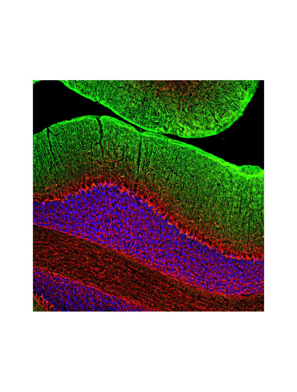
Image of a rat cerebellum section by Immunofluorescence and immunohistochemistry. The section was stained with R-1395-50, Neurofilament medium polypeptide (NF-M), Rabbit pAb, (red, 1:2,000) and co-stained with product M-1650-100, Growth associated protein 43 (GAP43), Mouse mAb, (green, 1:2,000). Method: Following transcardial perfusion of rat with 4% paraformaldehyde, brain was post-fixed for 24 hours, cut to 45 um, and free-floating sections were stained. The NF-M antibody strongly labels neuronal processes throughout the cerebellum, while the GAP43 antibody stains predominantly synaptic regions in the molecular layer.
Image of a rat cerebral cortex section by Immunofluorescence and Immunohistochemistry. The section was stained with R-1395-50, Neurofilament medium polypeptide (NF-M), Rabbit pAb, (red) and co-stained with a monoclonal antibody to the beta-adrendergic receptor kinase 1 (green). Product R-1395-50 reveals the perikarya of pyramidal neurons and dendrites and axons surrounding them.
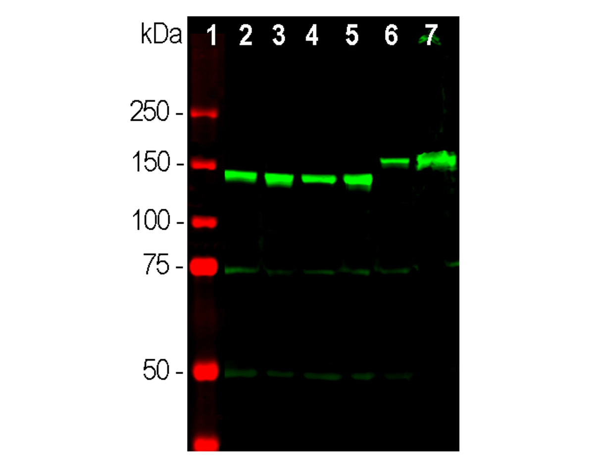
Analysis by western blot of NF-M expression in neuronal tissue lysates with R-1395-50, (green, 1:2,000) with strong bands at 145 kDa (rodent NF-M molecules) and 160 kDa (pig and other larger mammals including humans). Lane 1: protein standard; Lane 2: rat brain; Lane 3: rat spinal cord; Lane 4: mouse brain; Lane 5: mouse spinal cord; Lane 6: pig brain; Lane 7: pig spinal cord.
 1800 605-5127
 +61 (0)8 8352 7711