Alternative Names70 kDa lamin; Renal carcinoma antigen NY-REN-32; LMNA; LMN1;
Application(s)ICC, WB
Antibody HostChicken
Antibody TypePolyclonal
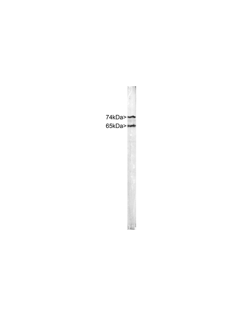
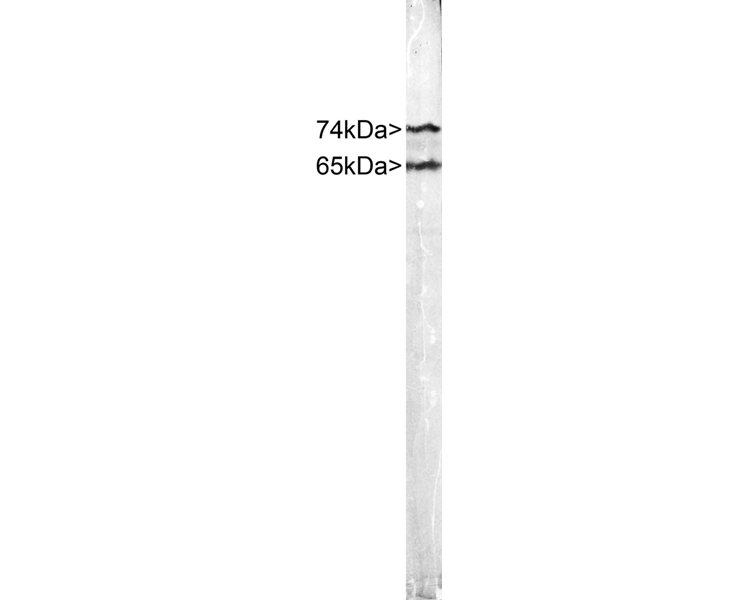
SpecificityLamin A and Lamin C. The antibody reacts with a 74 kDa and 65 kDa band by Western blot on HeLa cell extract. It has also been used successfully for immunocytochemistry on HeLa cell cultures.
Species ReactivityHuman
Immunogen DescriptionFull length recombinant human Lamin C
Application DetailsImmunocytochemistry (ICC) and Western Blotting (WB). A dilution of 1:1,000-1:2,000 is recommended for WB. A dilution of 1:500-1:1,000 is recommended for ICC. The optimal dilution should be determined by the end user.
TargetLamin A/C
SpecificityLamin A and Lamin C. The antibody reacts with a 74 kDa and 65 kDa band by Western blot on HeLa cell extract. It has also been used successfully for immunocytochemistry on HeLa cell cultures.
Target Host SpeciesHuman
Species ReactivityHuman
Antibody HostChicken
Antibody TypePolyclonal
Antibody IsotypeIgY
ConjugateUnconjugated
Immunogen DescriptionFull length recombinant human Lamin C
Purity DescriptionAffinity-purified
FormatLyophilized IgY preparation, with sodium azide.
Reconstitution InstructionsSpin vial briefly before opening. Reconstitute with 100 µL sterile-filtered, ultrapure water. Centrifuge to remove any insoluble material.
Storage InstructionsAfter reconstitution of lyophilized antibody, aliquot and store at -20°C for a higher stability. Avoid freeze-thaw cycles.
Batch NumberPlease see item label.
Expiration Date12 months after date of receipt (unopened vial).
Alternative Names70 kDa lamin; Renal carcinoma antigen NY-REN-32; LMNA; LMN1;
Scientific BackgroundThe Lamin proteins are members of the intermediate filament protein family but are located inside the nucleus rather than in the cytoplasm (1). The lamins function as skeletal components tightly associated with the inner nuclear membrane. Originally the proteins of the nuclear cytoskeleton were named Lamin A, B and C, from top to bottom as visualized on SDS-PAGE gels. Subsequently it was found that Lamins A and C were coded for by a single gene (2), while the Lamin B band may contain two proteins encoded by two genes now called Lamin B1 and Lamin B2. Lamin A has a mass of about 74 kDa while Lamin C is 65 kDa. The Lamin A protein includes 98 amino acids missing from Lamin C, while Lamin C has a C-terminal 6 amino acid peptide not present in Lamin A. Apart from these regions Lamin A and C are identical so that antibodies raised against either protein are likely to cross react with the other, as is the case with this monoclonal. Lamin polymerization and depolymerization is regulated by phosphorylation by cyclin dependent protein kinase 1 (CDK1), the key component of "maturation promoting factor", the central regulator of cell division. Activity of this kinase increases during cell division and is responsible for the breakdown of the nuclear lamina. Mutations in the LMNA gene are associated with several serious human diseases, including Emery-Dreifuss muscular dystrophy, familial partial lipodystrophy, limb girdle muscular dystrophy, dilated cardiomyopathy, Charcot-Marie-Tooth disease type 2B1, and Hutchinson-Gilford progeria syndrome. This family of diseases belong to a larger group which are often referred to as Laminopathies, though some laminopathies are associated in defects in Lamin B1, B2 or one or other of the numerous nuclear lamina binding proteins. A truncated version of lamin A, commonly known as progerin, causes Hutchinson-Gilford progeria syndrome, a form of premature aging (3).
Crude HeLa cell extract stained with Chicken anti-Lamin A/C. Two bands at 74 kDa and 65 kDa, corresponding to Lamin A and C.
HeLa cells stained with C-1698-100 (red), and counterstained with our monoclonal antibody to LAMP1 (M-1652-100). The Lamin A/C antibody reveals strong nuclear lamina staining, while the LAMP1 antibody reveals strong cytoplasmic punctate staining of lysosomes and early endosomes. The blue stain reveals DNA in the nuclei of these cells. Since both DNA (blue) and Lamin A/C (red) are associated with the nuclear compartment, this region appears crimson in this image.
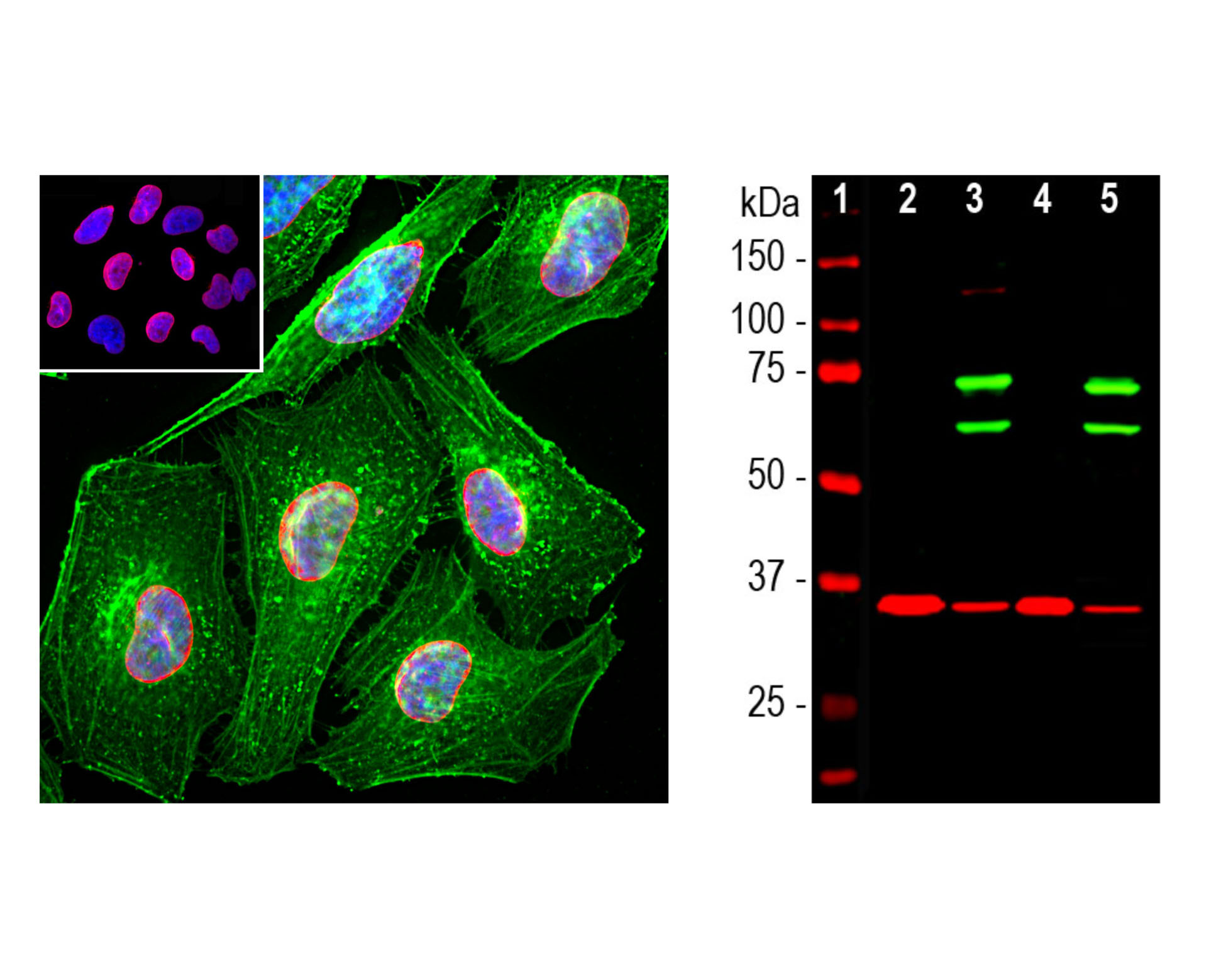
Left: Analysis of lamin A/C expression in HeLa cells with chicken anti-lamin A/C (red, 1:2,000), costained with mouse anti-actin (green) by Immunocytochemistry. Blue: Hoechst nuclear DNA stain. The chicken antibody specifically labels the nuclear lamina, while the actin antibody stains the submembranous actin-rich cytoskeleton, stress fibers and bundles of actin associated with cell adhesion sites. Right: Western blot analysis of cytosolic or nuclear enriched fractions of cell lines probed with chicken anti-lamin A/C (green, 1:1,000). [2] HeLa cytosol, [3] HeLa nuclear, [4] NIH-3T3 cytosol, and [5] NIH-3T3 nuclear fractions. Two strong bands at 65 kDa and 74 kDa correspond to lamin A and lamin C proteins, respectively, detected exclusively in the nuclear fractions. The same blot was simultaneously probed with mouse anti-GAPDH (M-1376-250, red). The single band at 37 kDa represents GAPDH protein
General ReferencesFisher, D. Z., Chaudhary, N., Blobel, G. cDNA sequencing of nuclear lamins A and C reveals primary and secondary structural homology to intermediate filament proteins. Proc. Nat. Acad. Sci. 83: 6450-6454 (1986). McKeon, F. D., Kirschner, M. W., Caput, D. Homologies in both primary and secondary structure between nuclear envelope and intermediate filament proteins. Nature 319: 463-468 (1986). Liu, B. and Zhou, Z. Lamin A/C, laminopathies and premature ageing. Histol. Histopathol. 23: 747-763 (2006).
 1800 605-5127
 +61 (0)8 8352 7711