 1800 605-5127
 +61 (0)8 8352 7711
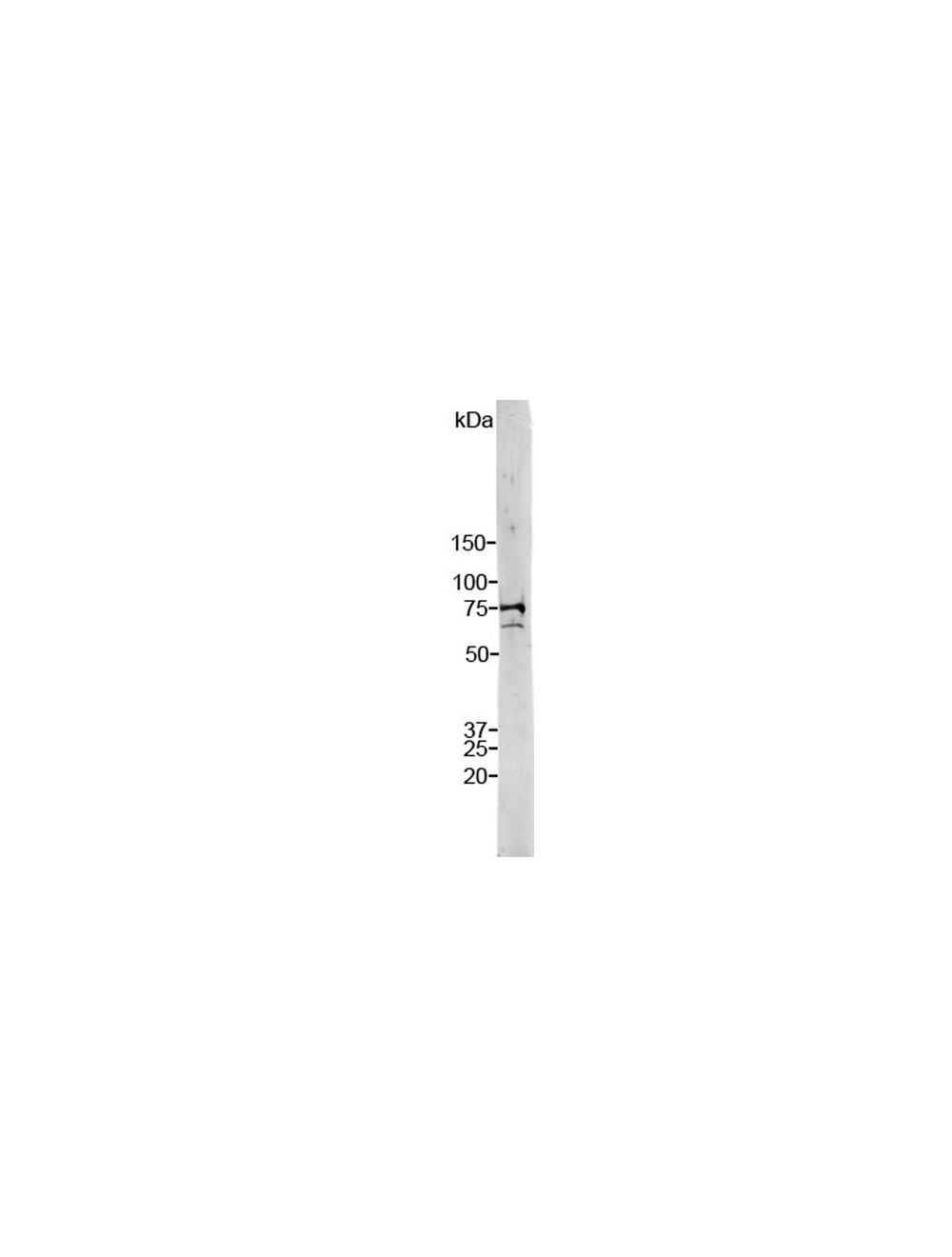

Lamin A/C, Rabbit Polyclonal Antibody
As low as
US$317.00
Only %1 left
Catalog Number
R-1697
- Product Name Lamin A/C, Rabbit Polyclonal Antibody
- Product Description Rabbit anti-Lamin A/C Polyclonal Antibody (Unconjugated), suitable for WB, ICC.
- Application(s) ICC, WB
- Antibody Host Rabbit
- Antibody Type Polyclonal
- Specificity Lamin A and Lamin C. The antibody reacts with a 74 kDa and 65 kDa band by Western blot on HeLa cell extract. It has also been used successfully for immunocytochemistry on HeLa cell cultures.
- Species Reactivity Human, Other Mammals (Predicted)
- Immunogen Description Full length recombinant human Lamin C
- Conjugate Unconjugated
- Purity Description Whole serum
- Regulatory Status For research use only.
Product Info
- Product Description Rabbit anti-Lamin A/C Polyclonal Antibody (Unconjugated), suitable for WB, ICC.
- Application(s) ICC, WB
- Application Details Immunocytochemistry (ICC) and Western Blotting (WB). A dilution of 1:1,000-1:5,000 is recommended for WB. A dilution of 1:1,000-1:5,000 is recommended for ICC. The optimal dilution should be determined by the end user.
- Target Lamin A/C
- Specificity Lamin A and Lamin C. The antibody reacts with a 74 kDa and 65 kDa band by Western blot on HeLa cell extract. It has also been used successfully for immunocytochemistry on HeLa cell cultures.
- Target Host Species Human
- Species Reactivity Human, Other Mammals (Predicted)
- Antibody Host Rabbit
- Antibody Type Polyclonal
- Antibody Isotype IgG
- Conjugate Unconjugated
- Immunogen Description Full length recombinant human Lamin C
- Purity Description Whole serum
- Format Lyophilized with sodium azide.
- Reconstitution Instructions Spin vial briefly before opening. Reconstitute with 100 µL sterile-filtered, ultrapure water. Centrifuge to remove any insoluble material.
- Storage Instructions After reconstitution of lyophilized antibody, aliquot and store at -20°C for a higher stability. Avoid freeze-thaw cycles.
- Batch Number Please see item label.
- Expiration Date 12 months after date of receipt (unopened vial).
- Uniprot Number P02545
- Uniprot Number/Name P02545 (LMNA_HUMAN)
- Scientific Background The Lamin proteins are members of the intermediate filament protein family but are located inside the nucleus rather than in the cytoplasm (1). The lamins function as skeletal components tightly associated with the inner nuclear membrane. Originally the proteins of the nuclear cytoskeleton were named Lamin A, B and C, from top to bottom as visualized on SDS-PAGE gels. Subsequently it was found that Lamins A and C were coded for by a single gene (2), while the Lamin B band may contain two proteins encoded by two genes now called Lamin B1 and Lamin B2. Lamin A has a mass of about 74 kDa while Lamin C is 65 kDa. The Lamin A protein includes 98 amino acids missing from Lamin C, while Lamin C has a C-terminal 6 amino acid peptide not present in Lamin A. Apart from these regions Lamin A and C are identical so that antibodies raised against either protein are likely to cross react with the other, as is the case with this monoclonal. Lamin polymerization and depolymerization is regulated by phosphorylation by cyclin dependent protein kinase 1 (CDK1), the key component of "maturation promoting factor", the central regulator of cell division. Activity of this kinase increases during cell division and is responsible for the breakdown of the nuclear lamina. Mutations in the LMNA gene are associated with several serious human diseases, including Emery-Dreifuss muscular dystrophy, familial partial lipodystrophy, limb girdle muscular dystrophy, dilated cardiomyopathy, Charcot-Marie-Tooth disease type 2B1, and Hutchinson-Gilford progeria syndrome. This family of diseases belong to a larger group which are often referred to as Laminopathies, though some laminopathies are associated in defects in Lamin B1, B2 or one or other of the numerous nuclear lamina binding proteins. A truncated version of lamin A, commonly known as progerin, causes Hutchinson-Gilford progeria syndrome, a form of premature aging (3).
- Shipping Temperature 25°C (ambient)
- UNSPSC CODE 41116161
- Regulatory Status For research use only.
Specifications


- General References Fisher, D. Z., Chaudhary, N., Blobel, G. cDNA sequencing of nuclear lamins A and C reveals primary and secondary structural homology to intermediate filament proteins. Proc. Nat. Acad. Sci. 83: 6450-6454 (1986).McKeon, F. D., Kirschner, M. W., Caput, D. Homologies in both primary and secondary structure between nuclear envelope and intermediate filament proteins. Nature 319: 463-468 (1986).Liu, B. and Zhou, Z. Lamin A/C, laminopathies and premature ageing. Histol. Histopathol. 23: 747-763 (2006).