Product DescriptionMouse anti-Microtubule Associated Protein 2 (MAP2) Monoclonal Antibody (Unconjugated), suitable for WB, IHC-Frozen, ICC.
Application(s)ICC, IHC-Frozen, WB
Application DetailsImmunohistochemistry (IHC), Immunocytochemistry (ICC) and Western Blotting (WB). A dilution of 1:1,000 - 1:5,000 is recommended for IHC and ICC, and 1:5,000-1:10,000 is recommended for WB. The optimal dilution should be determined by the end user.
TargetMicrotubule Associated Protein 2 (MAP2)
SpecificityThe specificity of this antibody has been confirmed by WB and IHC against the antigen. Human; Rat; Mouse;
Target Host SpeciesBovine
Species ReactivityBovine, Human, Mouse, Rat
Antibody HostMouse
Antibody TypeMonoclonal
Antibody IsotypeIgG
Clone Name5H11
ConjugateUnconjugated
Immunogen DescriptionHigh molecular MAP protein preparation derived from bovine brain
Purity DescriptionProtein G purified
FormatLyophilized from PBS buffer pH 7.2-7.6 with 0.1% trehalose, and sodium azide
Reconstitution InstructionsSpin vial briefly before opening. Reconstitute with 100 µL sterile-filtered, ultrapure water to achieve a 1 mg/mL concentration. Centrifuge to remove any insoluble material.
Storage InstructionsAt least 12 months after purchase at 2-8°C (lyophilized formulations). After reconstitution, aliquot and store at -20°C for a higher stability. Avoid freeze-thaw cycles.
Batch NumberPlease see item label.
Expiration Date12 months after date of receipt (unopened vial).
Alternative NamesMicrotubule-associated protein 2; MAP-2; Mtap2;
Scientific BackgroundMicrotubules are 25nm diameter protein rods found in most kinds of eukaryotic cells. They are polymerized from a dimeric subunit made of one 'a' subunit and one 'b' tubulin subunit. Microtubules are associated with a family of proteins called microtubule associated proteins (MAPs), which includes the protein t (tau) and a group of proteins referred to as MAP1, MAP2, MAP3, MAP4 and MAP5. MAP2 is made up of two ~280 kDa apparent molecular weight bands referred to as MAP2 a and MAP2 b. A third lower molecular weight form, usually called MAP2c, corresponds to a pair of protein bands running at ~70 kDa on SDS-PAGE gels. All these MAP2 forms are derived from a single gene by alternate transcription, and all share a C-terminal sequence which includes either three or four microtubule binding peptide sequences, which are very similar to those found in the related microtubule binding protein t (tau). MAP2 isoforms are expressed only in neuronal cells and specifically in the perikarya and dendrites of these cells. Antibodies to MAP2 are therefore excellent markers on neuronal cells, their perikarya and neuronal dendrites.
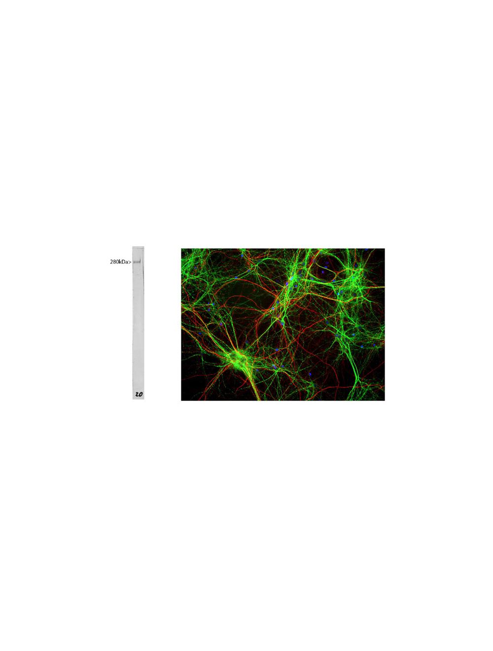
Whole rat brain lysate with mouse anti-MAP2 antibody. The antibody recognizes the ~280 kDa protein. Right: Mixed neuron/glia cultures stained with mouse anti-MAP2 (green) and also rabbit antibody to neurofilament H (Catalog Number R-1388-50) (red). Since the NF-H protein is largely expressed in neuronal axons, while the MAP2 is only found in neuronal dendrites and perikarya, there is little overlap between these two staining patterns. DNA stain shows nuclei of neurons and non-neuronal cells (blue).
Left: Rat hippocampus section stained by Immunohistochemistry with mouse anti-MAP2 (green, 1:5,000) and rabbit antibody to alpha-internexin (R-1379-50, red, 1:2,000). IHC Method: Following transcardial perfusion of rat with 4% paraformaldehyde, brain was post-fixed for 24 hours, cut to 45 um, and free-floating sections were stained. MAP2 protein is detected in the perikarya and dendrites of the most neurons, and the alpha-internexin antibody selectively stains axons and dendrites of neuronal cells. Right: Western blot analysis of tissue lysates using mouse anti-MAP2 (green, 1:10,000). [1] protein standard, [2] adult rat whole brain, [2] embryonic (E20) rat brain, [4] adult rat spinal cord, and [5] adult mouse brain. A band at about 280 kDa corresponds to full length MAP2A and MAP2B protein. MAP2A/B is expressed heavily in adult brain particularly in cortical regions, but is a more minor component of spinal cord and almost absent from the embryonic brain sample. Note that the epitope for this antibody in within the projection domain found only in MAP2A and MAP2B, thus the antibody does not bind to the lower molecular weight MAP2C and MAP2D isoforms which lack this region.
MAP2 staining in fixed and permeabilized striatal neurons (7 DIV) by Immunocytochemistry. Primary antibody concentration: 1 µg/mL. Image courtesy of QBM Cell Science.
 1800 605-5127
 +61 (0)8 8352 7711