 1800 605-5127
 +61 (0)8 8352 7711
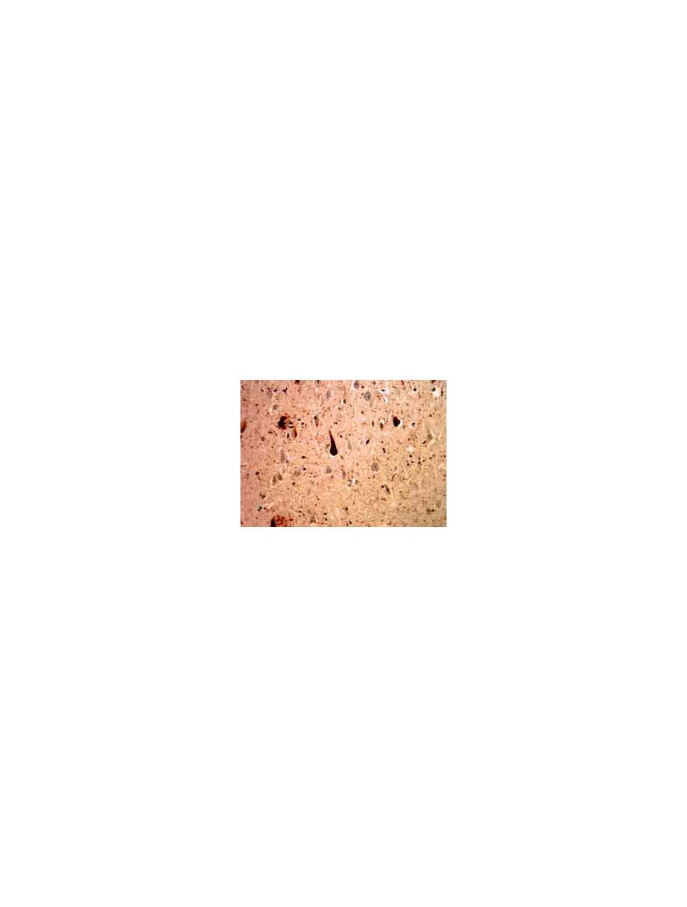

Polyubiqutin-B, Mouse Monoclonal Antibody
- Product Name Polyubiqutin-B, Mouse Monoclonal Antibody
- Product Description Mouse anti-Polyubiqutin-B Monoclonal Antibody (Unconjugated), suitable for WB, IHC-Frozen, IHC-Paraffin-embedded.
- Alternative Names RPS27A; UBA52; UBB; UBC; Polyubiquitin-B; Polyubiquitin-C;
- Application(s) IHC-Frozen, IHC-Paraffin-embedded, WB
- Antibody Host Mouse
- Antibody Type Monoclonal
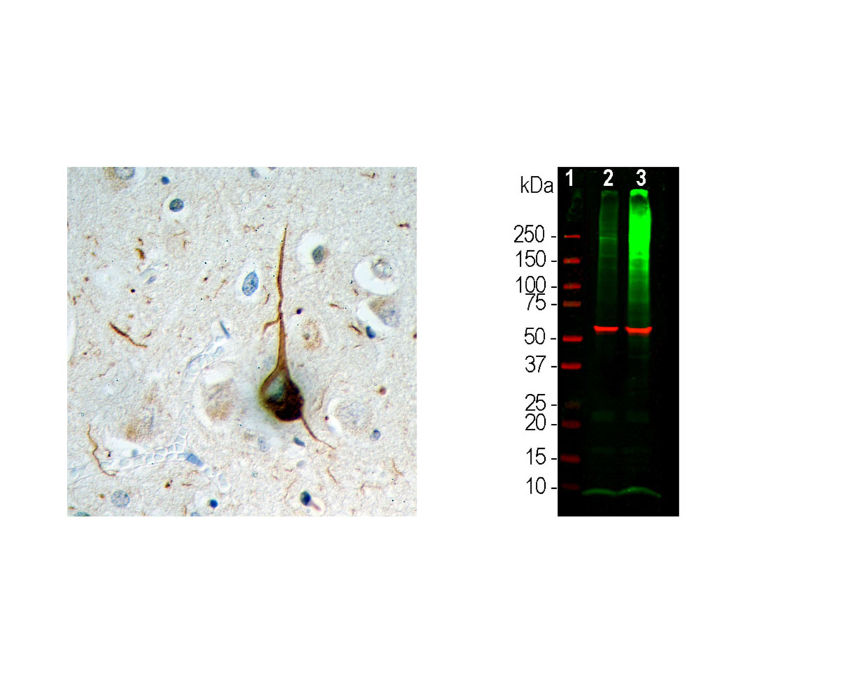
- Specificity The specificity of this antibody has been confirmed by WB. This antibody detects ~8.5 kDa Ubiquitin. Hu, Bov, Chk, Drosophila, and C. elegans
- Species Reactivity Bovine, C. elegans, Chicken, Drosophila, Human
- Immunogen Description Raised against purified ubiquitin conjugated with glutaraldehyde to keyhole limpet hemocyanin.
- Conjugate Unconjugated
- Purity Description Protein G purified
- Regulatory Status For research use only.
Product Info
- Product Description Mouse anti-Polyubiqutin-B Monoclonal Antibody (Unconjugated), suitable for WB, IHC-Frozen, IHC-Paraffin-embedded.
- Application(s) IHC-Frozen, IHC-Paraffin-embedded, WB
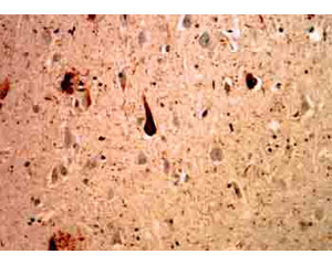
- Application Details Western Blotting (WB), Immunohistochemistry - paraffin embedded tissue (IH-P) and ELISA. Suggested dilution for WB is 1:500-1,000. This antibody can be used on mildly fixed histological sections of human brain for studies of Alzheimer's disease. This antibody also works on paraffin embedded material. It also recognises other ubiquinated inclusion bodies such as Lewy bodies of Parkinson's disease and the Pick bodies in Pick's disease in formalin fixed tissues. Suggested dilution for IH is 1:500. Biosensis recommends optimal dilutions/concentrations should be determined by the end user.
- Target Polyubiqutin-B
- Specificity The specificity of this antibody has been confirmed by WB. This antibody detects ~8.5 kDa Ubiquitin. Hu, Bov, Chk, Drosophila, and C. elegans
- Target Host Species Human
- Species Reactivity Bovine, C. elegans, Chicken, Drosophila, Human
- Antibody Host Mouse
- Antibody Type Monoclonal
- Antibody Isotype IgG1
- Clone Name Ubi-1
- Conjugate Unconjugated
- Immunogen Description Raised against purified ubiquitin conjugated with glutaraldehyde to keyhole limpet hemocyanin.
- Purity Description Protein G purified
- Format Lyophilized from PBS buffer pH 7.2-7.6 with 0.1% trehalose, and sodium azide
- Reconstitution Instructions Spin vial briefly before opening. Reconstitute with 100 µL sterile-filtered, ultrapure water to achieve a 1 mg/mL concentration. Centrifuge to remove any insoluble material.
- Storage Instructions After reconstitution of lyophilized antibody, aliquot and store at -20°C for a higher stability. Avoid freeze-thaw cycles.
- Batch Number Please see item label.
- Expiration Date 12 months after date of receipt (unopened vial).
- Alternative Names RPS27A; UBA52; UBB; UBC; Polyubiquitin-B; Polyubiquitin-C;
- Uniprot Number P0CG47
- Uniprot Number/Name P0CG47 (UBB_HUMAN)
- Scientific Background Ubiquitin is a highly conserved 76 amino acid protein with an estimated molecular weight of 8.56 kDa which has a central role in regulated protein degradation. It is a protein modifier which can be covalently attached to target lysines either as a monomer or as a lysine-linked polymer. Several types of polymeric chains can be formed depending on the lysine used for the assembly. Attachment to proteins as a polymer leads to their degradation by the 26S proteosome; a complex, multicatalytic cytosolic and nuclear protease. Attachment to proteins as a monomer or as an alternatively linked polymer does not lead to proteasomal degradation and may be required for numerous functions, including maintenance of chromatic structure, regulation of gene expression, stress response, ribosome biogenesis and DNA repair. Ubiquitin is synthesized as a polyubiquitin precursor with exact head to tail repeats, the number of repeats of which differ between species and strains. In some species there is a final amino-acid after the last repeat, here in bovine a Cys. Some ubiquitin genes contain a single copy of ubiquitin fused to a ribosomal protein (either L40 or S27a).
- Shipping Temperature 25°C (ambient)
- UNSPSC CODE 41116161
- Regulatory Status For research use only.
Specifications
-
Specific References
Josephs K.A. et al (2006) Atypical progressive supranuclear palsy with corticospinal tract degeneration. J Neuropathol Exp Neurol. 2006 Apr;65(4):396-405.
Josephs K.A. et al (2007) Neuropathologic features of frontotemporal lobar degeneration with ubiquitin-positive inclusions with progranulin gene (PGRN) mutations. J Neuropathol Exp Neurol. 2007 Feb;66(2):142-51.
Rudzinski L.A. et al (2008) Early onset familial Alzheimer Disease with spastic paraparesis, dysarthria, and seizures and N135S mutation in PSEN1. Alzheimer Dis Assoc Disord. 2008 Jul-Sep;22(3):299-307.
Josephs K.A. et al (2009) Evaluation of subcortical pathology and clinical correlations in FTLD-U subtypes. Acta Neuropathol. 2009 Sep;118(3):349-58.