Alternative NamesGamma-aminobutyric acid receptor-associated protein-like 2; GABA(A) receptor-associated protein-like 2; Ganglioside expression factor 2; GEF-2; General protein transport factor p16; MAP1 light chain 3-related protein; GABARAPL2; FLC3A; GEF2;
Application(s)IHC-Frozen, WB
Antibody HostRabbit
Antibody TypePolyclonal
SpecificityIHC, WB and ELISA confirmed the specificity for GABARAP L2 (GABARAPL2). A 14 kDa band, that corresponds to the molecular weight of GABARAPL2, is detected via western blot analysis. Human, rat. Other species not yet tested.
Species ReactivityHuman, Rat
Immunogen DescriptionA synthetic peptide (CVESAKIRAKYP) corresponding to the N-terminal of human GABARAP L2 (GABARAPL2) protein has been used as the immunogen. The sequence is homologous with mouse and rat form of GABARAP L2 (GABARAPL2).
Application DetailsIHC, immunofluorescence, WB. A dilution of 1:200 to 1:1000 dilution is recommended for these applications. Biosensis recommends optimal dilutions/concentrations should be determined by the end user.
SpecificityIHC, WB and ELISA confirmed the specificity for GABARAP L2 (GABARAPL2). A 14 kDa band, that corresponds to the molecular weight of GABARAPL2, is detected via western blot analysis. Human, rat. Other species not yet tested.
Target Host SpeciesHuman
Species ReactivityHuman, Rat
Antibody HostRabbit
Antibody TypePolyclonal
Antibody IsotypeMixed
ConjugateUnconjugated
Immunogen DescriptionA synthetic peptide (CVESAKIRAKYP) corresponding to the N-terminal of human GABARAP L2 (GABARAPL2) protein has been used as the immunogen. The sequence is homologous with mouse and rat form of GABARAP L2 (GABARAPL2).
Purity DescriptionWhole serum
FormatLyophilized
Reconstitution InstructionsSpin vial briefly before opening. Reconstitute in 100 µL sterile-filtered, ultrapure water. Centrifuge to remove any insoluble material.
Storage InstructionsAfter reconstitution keep aliquots at -20°C for a higher stability, and at 2-8°C with an appropriate antibacterial agent. Glycerol (1:1) may be added for an additional stability. Avoid repetitive freeze/thaw cycles.
Batch NumberPlease see item label.
Expiration Date12 months after date of receipt (unopened vial).
Alternative NamesGamma-aminobutyric acid receptor-associated protein-like 2; GABA(A) receptor-associated protein-like 2; Ganglioside expression factor 2; GEF-2; General protein transport factor p16; MAP1 light chain 3-related protein; GABARAPL2; FLC3A; GEF2;
Scientific BackgroundFUNCTION: Involved in intra-Golgi traffic. Modulates intra-Golgi transport through coupling between NSF activity and SNAREs activation. It first stimulates the ATPase activity of NSF which in turn stimulates the association with GOSR1. SUBUNIT: Monomer. Interacts with GABRG2, NSF, GOSR1 and beta-tubulin. Interacts with ULK1. SUBCELLULAR LOCATION: Golgi apparatus. TISSUE SPECIFICITY: Ubiquitous. Expressed at high levels in the brain, heart, prostate, ovary, spleen and skeletal muscle. Expressed at very low levels in lung, thymus and small intestine. SIMILARITY: Belongs to the MAP1 LC3 family. ESTIMATED MOLECULAR WEIGHT: 13.667 kDa.
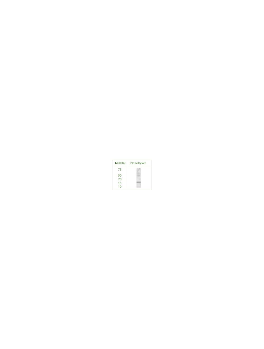
Western blot under reducing conditions on 293 cell lysate using Rabbit antibody to GABARAP L2 (GABARAPL2): whole serum (R-142-100) at a dilution of 1:100.
A. Confocal microscopy on immunofluorescently detected MPO (in green) in cytospin-isolated human white blood cells counter-stained with Hoechst Dye. Here, the merged picture is presented.
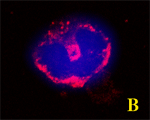
B. Confocal microscopy on immunofluorescently detected GABARAP L2 (GABARAPL2) in cytospin-isolated human white blood cells using Rabbit antibody to GABARAP L2 (GABARAPL2): whole serum (R-142-100) at a dilution of 1: 200, incubated for 1 h at room temperature. The cells were stained for GABARAP L2 (GABARAPL2) appearing in red counter-stained with Hoechst DyeHere. The merged picture is presented.
C is the merged picture of Figures A & B. Confocal microscopy on immunofluorescently detected GABARAP L2 (GABARAPL2) in cytospin-isolated human white blood cells using Rabbit antibody to GABARAP L2 (GABARAPL2): whole serum (R-142-100) at a dilution of 1: 200, incubated for 1 h at room temperature. The cells were double-stained for GABARAP L2 (GABARAPL2) appearing in red and Myeloperoxidase (MPO) appearing in green. The cells were counter stained with Hoechst Dye (blue colour).
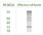
Western blot under reducing conditions on Odora (an Olfactory cell line) cell lysate using Rabbit antibody to GABARAP L2 (GABARAPL2): whole serum (R-142-100) at a dilution of 1:100.
General ReferencesOkazaki N. et al. Brain Res. Mol. Brain Res. 85:1-12(2000) Xin Y. et al. Genomics 74:408-413(2001) The MGC Project Team. Genome Res. 14:2121-2127(2004)
 1800 605-5127
 +61 (0)8 8352 7711