SpecificityIHC, WB and ELISA confirmed the specificity for GABARAP. Human, rat. Other species not yet tested.
Species ReactivityHuman, Rat
Immunogen DescriptionA synthetic peptide (FEKRRSEGEKIC) corresponding to the N-terminal of human GABARAP protein has been used as the immunogen. The sequence is homologous with mouse and rat form of GABARAP.
Product DescriptionRabbit anti-Gamma-aminobutyric acid receptor-associated protein (GABARAP) Polyclonal Antibody (Unconjugated), suitable for WB, IHC-Frozen.
Application(s)IHC-Frozen, WB
Application DetailsIHC, immunofluorescence, WB. A dilution of 1:200 to 1:1000 dilution is recommended for these applications. Biosensis recommends optimal dilutions/concentrations should be determined by the end user.
TargetGamma-aminobutyric acid receptor-associated protein (GABARAP)
SpecificityIHC, WB and ELISA confirmed the specificity for GABARAP. Human, rat. Other species not yet tested.
Target Host SpeciesHuman
Species ReactivityHuman, Rat
Antibody HostRabbit
Antibody TypePolyclonal
Antibody IsotypeMixed
ConjugateUnconjugated
Immunogen DescriptionA synthetic peptide (FEKRRSEGEKIC) corresponding to the N-terminal of human GABARAP protein has been used as the immunogen. The sequence is homologous with mouse and rat form of GABARAP.
Purity DescriptionWhole serum
FormatLyophilized
Reconstitution InstructionsSpin vial briefly before opening. Reconstitute in 100 µL sterile-filtered, ultrapure water. Centrifuge to remove any insoluble material.
Storage InstructionsAfter reconstitution keep aliquots at -20°C for a higher stability, and at 2-8°C with an appropriate antibacterial agent. Glycerol (1:1) may be added for an additional stability. Avoid repetitive freeze/thaw cycles.
Batch NumberPlease see item label.
Expiration Date12 months after date of receipt (unopened vial).
Scientific BackgroundGABARAP is highly positively charged in its N-terminus and shares sequence homology with MAP1LC3 1A and 1B. This protein clusters neurotransmitter receptors (GABA(A) receptors) by mediating interaction with the cytoskeleton. SUBUNIT: Interacts with GABRG2, TUBA1, ULK1 and NSF. Interacts with beta-tubulin and GPHN. SUBCELLULAR LOCATION: Intracytoplasmic membrane. Cytoskeleton. Largely associated with intracellular membrane structures including the Golgi apparatus and post-synaptic cisternae. Colocalizes with microtubules. TISSUE SPECIFICITY: Heart, brain, placenta, skeletal muscle, kidney and pancreas.
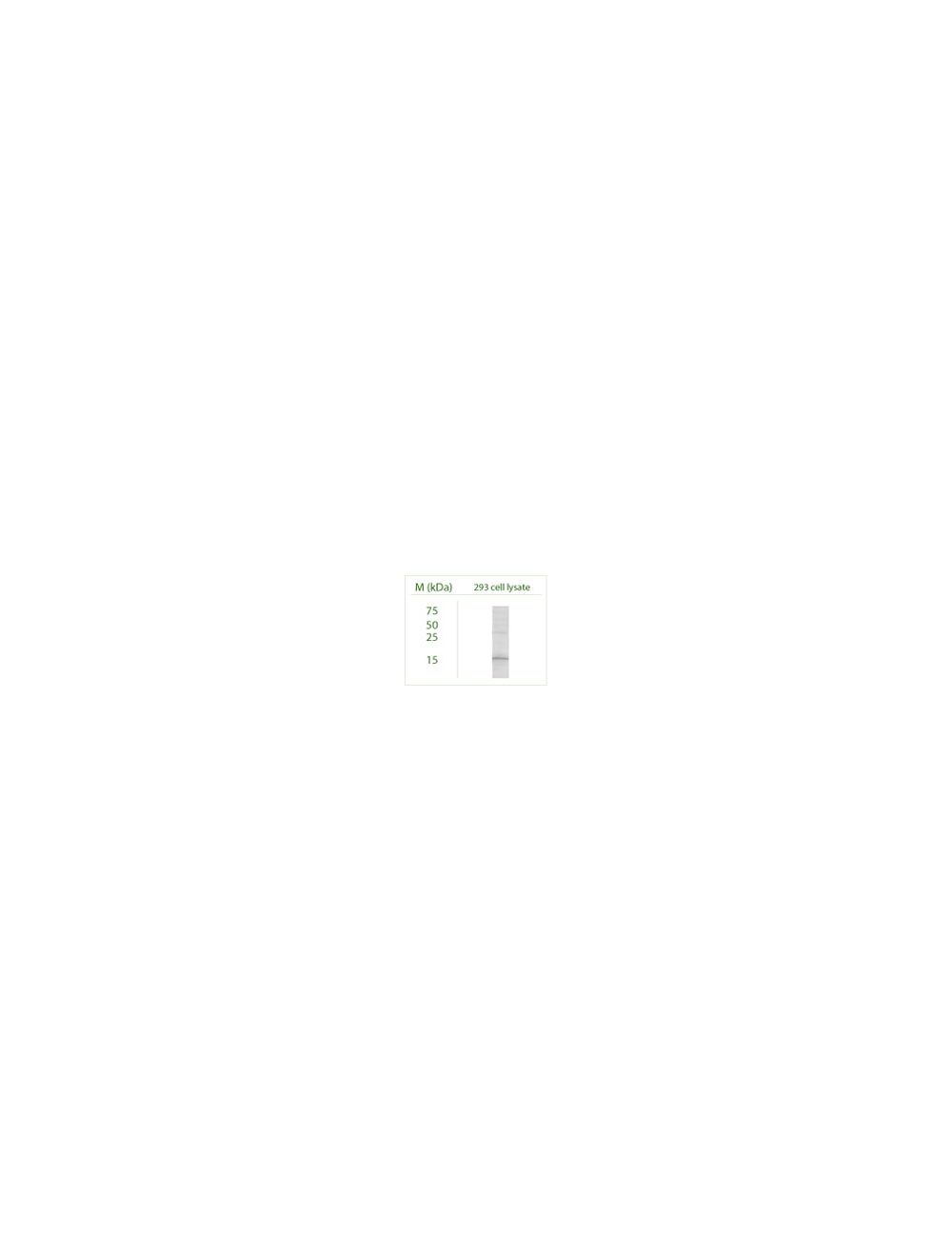
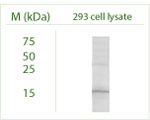
Western blot under reducing conditions on 293 cell lysate using Rabbit antibody to GABARAP: whole serum (R-143-100) at a dilution of 1:100.
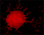
A. Confocal microscopy on immunofluorescently detected GABARAP (in red) in cytospin-isolated human white blood cells using Rabbit antibody to GABARAP: whole serum (R-143-100) at a dilution of 1: 200, incubated for 1 h at room temperature, counter-stained with Hoechst Dye. Here, the merged picture is presented.
B. Confocal microscopy on immunofluorescently detected MHC class II (in green) in cytospin-isolated human white blood cells. The cells were counter-stained with Hoechst Dye. The merged picture is presented.
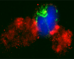
C is the merged picture of Figures A & B. Confocal microscopy on immunofluorescently detected GABARAP in cytospin-isolated human white blood cells using Rabbit antibody to GABARAP: whole serum (R-143-100) at a dilution of 1: 200, incubated for 1 h at room temperature. The cells were double-stained for GABARAP appearing in red and MHC class II appearing in green. The cells were counter stained with Hoechst Dye (blue colour). Here, the merged picture is presented.
Confocal microscopy on immunofluorescently detected GABARAP (in red) in cytospin-isolated human white blood cells using Rabbit antibody to GABARAP: whole serum (R-143-100) at a dilution of 1: 200, incubated for 1 h at room temperature.
Confocal microscopy on immunofluorescently detected GABARAP in cytospin-isolated human white blood cells using Rabbit antibody to GABARAP: whole serum (R-143-100) at a dilution of 1: 200, incubated for 1 h at room temperature. The cells were double-stained for GABARAP appearing in red and MHC class II appearing in green. The cells were counter stained with Hoechst Dye (blue colour). Here, the merged picture is presented.
Confocal microscopy on immunofluorescently detected GABARAP in cytospin-isolated human white blood cells using Rabbit antibody to GABARAP: whole serum (R-143-100) at a dilution of 1: 200, incubated for 1 h at room temperature. The cells were double-stained for GABARAP appearing in red and MHC class II appearing in green. The cells were counter stained with Hoechst Dye (blue colour). Here, the merged picture is presented.
Western blot under reducing conditions on Odora (an Olfactory cell line) cell lysate using Rabbit antibody to GABARAP: whole serum (R-143-100) at a dilution of 1:100.
General ReferencesWang H. et al. Nature. 397: 69-72 (1999) Okazaki N. et al. Brain Res. Mol. Brain Res. 85:1-12(2000) Hu R.-M. et al. Proc. Natl. Acad. Sci. U.S.A. 97:9543-9548(2000) Knight D. et al. Biol. Chem. 277:5556-5561(2002)
 1800 605-5127
 +61 (0)8 8352 7711