Product DescriptiongoogleRabbit anti-Microtubule-associated protein 1 light chain 3 gamma (MAP1LC3C) Polyclonal Antibody (Unconjugated), suitable for WB, IHC-Frozen, ICC.
Alternative NamesMicrotubule-associated proteins 1A/1B light chain 3C; Microtubule-associated protein 1 light chain 3 gamma; MAP1A/MAP1B LC3 C; MAP1A/1B light chain 3 C; MAP1 light chain 3-like protein 3; Autophagy-related protein LC3 C; Autophagy-related ubiquitin-like modifier LC3 C; APG8c; MAP1LC3C
Application(s)ICC, IHC-Frozen, WB
Antibody HostRabbit
Antibody TypePolyclonal
SpecificityIHC and WB confirmed the specificity for MAP1LC3 C . This antibody should recognise MAP1LC3 C only and not the other forms MAP1LC3A and MAP1LC3B. Human, other species have not yet been tested.
Species ReactivityHuman
Immunogen DescriptionA synthetic peptide (CQEEVAGIRAKF) corresponding to the N-terminal of human MAP1LC3 C conjugated to Blue Carrier Protein has been used as the immunogen. The peptide is homologous with the corresponding sequence deriven from MAP1LC3 C protein in Macaca mulatta (monkey) and Canis familiaris (dog).
Product DescriptionRabbit anti-Microtubule-associated protein 1 light chain 3 gamma (MAP1LC3C) Polyclonal Antibody (Unconjugated), suitable for WB, IHC-Frozen, ICC.
Application(s)ICC, IHC-Frozen, WB
Application DetailsIHC, immunofluorescence, WB. A dilution of 1:100 to 1:1000 dilution is recommended for these applications. Biosensis recommends optimal dilutions/concentrations should be determined by the end user.
TargetMicrotubule-associated protein 1 light chain 3 gamma (MAP1LC3C)
SpecificityIHC and WB confirmed the specificity for MAP1LC3 C . This antibody should recognise MAP1LC3 C only and not the other forms MAP1LC3A and MAP1LC3B. Human, other species have not yet been tested.
Target Host SpeciesHuman
Species ReactivityHuman
Antibody HostRabbit
Antibody TypePolyclonal
Antibody IsotypeMixed
ConjugateUnconjugated
Immunogen DescriptionA synthetic peptide (CQEEVAGIRAKF) corresponding to the N-terminal of human MAP1LC3 C conjugated to Blue Carrier Protein has been used as the immunogen. The peptide is homologous with the corresponding sequence deriven from MAP1LC3 C protein in Macaca mulatta (monkey) and Canis familiaris (dog).
Purity DescriptionWhole serum
FormatLyophilized
Reconstitution InstructionsSpin vial briefly before opening. Reconstitute in 100 µL sterile-filtered, ultrapure water. Centrifuge to remove any insoluble material.
Storage InstructionsAfter reconstitution keep aliquots at -20°C for a higher stability, and at 2-8°C with an appropriate antibacterial agent. Glycerol (1:1) may be added for an additional stability. Avoid repetitive freeze/thaw cycles.
Batch NumberPlease see item label.
Expiration Date12 months after date of receipt (unopened vial).
Alternative NamesMicrotubule-associated proteins 1A/1B light chain 3C; Microtubule-associated protein 1 light chain 3 gamma; MAP1A/MAP1B LC3 C; MAP1A/1B light chain 3 C; MAP1 light chain 3-like protein 3; Autophagy-related protein LC3 C; Autophagy-related ubiquitin-like modifier LC3 C; APG8c; MAP1LC3C
Scientific BackgroundFUNCTION: Probably involved in formation of autophagosomal vacuoles (autophagosomes). SUBUNIT: 3 different light chains, LC1, LC2 and LC3, can associate with MAP1A and MAP1B proteins. SUBCELLULAR LOCATION: LC3-I: Cytoplasm. LC3-II: Intracytoplasmic membrane; lipid-anchor. LC3-II binds to the autophagic membranes. TISSUE SPECIFICITY: Most abundant in placenta, lung and ovary. PTM: The precursor molecule is cleaved by APG4B/ATG4B to form the cytosolic form, LC3-I. This is activated by APG7L/ATG7, transferred to ATG3 and conjugated to phospholipid to form the membrane-bound form, LC3-II. SIMILARITY: Belongs to the MAP1 LC3 family.
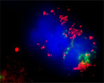
Confocal microscopy on immunofluorescently detected MAP1LC3 C in cytospin-isolated human white blood cells using Rabbit antibody to MAP1LC3 C : whole serum (R-140-100) at a dilution of 1: 200, incubated for 1 h at room temperature. The detected MAP1LC3 C appears red. The cells were also stained for Myeloperoxidase (MPO) appearing in green. The cells were counter stained with Hoechst Dye(blue colour). Here, the merged picture is presented.
Confocal microscopy on immunofluorescently detected MAP1LC3 C in cytospin-isolated human white blood cells using Rabbit antibody to MAP1LC3 C : whole serum (R-140-100) at a dilution of 1: 200, incubated for 1 h at room temperature. The detected MAP1LC3 C appears red. The cells were also stained for Myeloperoxidase (MPO) appearing in green. The cells were counter stained with Hoechst Dye (blue colour). Here, the merged picture is presented.
Confocal microscopy on immunofluorescently detected MAP1LC3 C in cytospin-isolated human white blood cells using Rabbit antibody to MAP1LC3 C : whole serum (R-140-100) at a dilution of 1: 200, incubated for 1 h at room temperature. The detected MAP1LC3 C appears red. The cells were also stained for Myeloperoxidase (MPO) appearing in green. The cells were counter stained with Hoechst Dye (blue colour). Here, the merged picture is presented.
Confocal microscopy on immunofluorescently detected MAP1LC3 C in cytospin-isolated human white blood cells using Rabbit antibody to MAP1LC3 C : whole serum (R-140-100) at a dilution of 1: 200, incubated for 1 h at room temperature. The detected MAP1LC3 C appears red. The cells were also stained for Myeloperoxidase (MPO) appearing in green. The cells were counter stained with Hoechst Dye (blue colour). Here, the merged picture is presented.
Western blot under reducing conditions on 293 cell lysate using Rabbit antibody to MAP1LC3 C : whole serum (R-140-100) at a dilution of 1:100.
General ReferencesGreenberg JT. Dev Cell. 8(6):799-80(2005) Baehrecke EH. Nat Rev Mol Cell Biol. 6(6):505-(2005) Levine B. Cell. 120(2):159-6(2005) Tanida I., et al. J. Biol. Chem. 279:36268-36276(2004) Lum JJ, et al. Nat Rev Mol Cell Biol. 6(6):439-4(2005) Tanida I., et al. Int. J. Biochem. Cell Biol. 36:2503-2518(2004)
 1800 605-5127
 +61 (0)8 8352 7711