Alternative NamesMicrotubule-associated proteins 1A/1B light chain 3A; MAP1A/MAP1B LC3 A; MAP1A/1B light chain 3 A; MAP1 light chain 3-like protein 1; Microtubule-associated protein 1 light chain 3 alpha; Autophagy-related protein LC3 A; Autophagy-related ubiquitin-like modifier LC3 A; APG8a; MAP1LC3A
Application(s)ICC, WB
Antibody HostRabbit
Antibody TypePolyclonal
SpecificityIHC, WB and ELISA confirmed the specificity for MAP1LC3 A. Human, rat. Other species not yet tested.
Species ReactivityHuman, Rat
Immunogen DescriptionA synthetic peptide (RSFADRCKEVQQI) corresponding to the N-terminal of human MAP1LC3 A protein conjugated to Blue Carrier Protein has been used as the immunogen. The sequence is homologous with mouse and rat MAP1LC3 A.
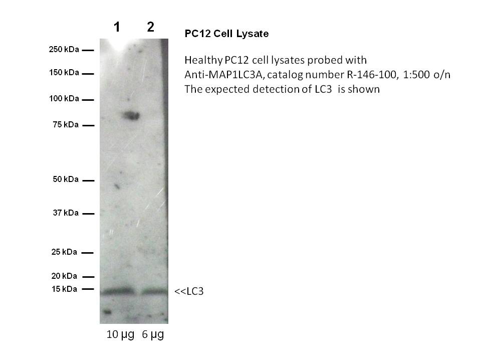
Application DetailsIHC, immunohistochemistry on 4% PFA fixed or 2% PLP fixation, 1:100-1000 primary antibody dilution. IC, Immunocytochemistry: acetone fixed specimens recommended. 1:100-1:1000 primary antibody dilution. Western Blot: R-146-100 requires samples to be denatured ONLY by boiling in SDS solution, not reduced. No signal is achieved using reduced samples. (Samples treated with DTT or Beta mercaptoethanol). 1:500 overnight 4 degrees is recommended for primary antibody dilution in western blots. Biosensis recommends optimal dilutions/concentrations should be determined by the end user.
SpecificityIHC, WB and ELISA confirmed the specificity for MAP1LC3 A. Human, rat. Other species not yet tested.
Target Host SpeciesHuman
Species ReactivityHuman, Rat
Antibody HostRabbit
Antibody TypePolyclonal
Antibody IsotypeMixed
ConjugateUnconjugated
Immunogen DescriptionA synthetic peptide (RSFADRCKEVQQI) corresponding to the N-terminal of human MAP1LC3 A protein conjugated to Blue Carrier Protein has been used as the immunogen. The sequence is homologous with mouse and rat MAP1LC3 A.
Purity DescriptionWhole serum
FormatLyophilized
Reconstitution InstructionsSpin vial briefly before opening. Reconstitute in 100 µL sterile-filtered, ultrapure water. Centrifuge to remove any insoluble material.
Storage InstructionsAfter reconstitution keep aliquots at -20°C for higher stability, and at 2-8°C with an appropriate antibacterial agent. Glycerol (1:1) may be added for an additional stability. Avoid repetitive freeze/thaw cycles.
Batch NumberPlease see item label.
Expiration Date12 months after date of receipt (unopened vial).
Alternative NamesMicrotubule-associated proteins 1A/1B light chain 3A; MAP1A/MAP1B LC3 A; MAP1A/1B light chain 3 A; MAP1 light chain 3-like protein 1; Microtubule-associated protein 1 light chain 3 alpha; Autophagy-related protein LC3 A; Autophagy-related ubiquitin-like modifier LC3 A; APG8a; MAP1LC3A
Scientific BackgroundMAP1A and MAP1B are microtubule-associated protein which mediate the physical interactions between microtubules and components of the cytoskeleton (probably involved in autophagosome formation). MAP1A and MAP1B each consist of a heavy chain subunit and 3 different light chain subunits (LC1, LC2 and LC3). MAP1LC3A is one of the light chain subunits and can associate with either MAP1A or MAP1B. The precursor form of MAP1LC3A is cleaved by APG4/ATG4B to form the cytosolic form LC3-1. This is activated by APG7L/ATG7, transferred to ATG3 and conjugated to phospholipid to form the membrane-bound form, LC3-II. MAP1LC3A is most abundant in heart, brain, liver, skeletal muscle and testis but is absent in thymus and peripheral leukocytes.
Confocal microscopy on immunofluorescently detected MAP1LC3 A in cytospin-isolated human white blood cells using Rabbit antibody to MAP1LC3 A : whole serum (R-146-100) at a dilution of 1: 200, incubated for 1 h at room temperature. The cells were double-stained for MAP1LC3 A appearing in red and MHC class II appearing in. The cells were counter stained with Hoechst Dye (blue colour). Here, the merged picture is presented.
PC12 non-reduced samples (boiled in SDS only, no DTT or BME)
A. Confocal microscopy on immunofluorescently detected MHC class II in cytospin-isolated human white blood cells.
B. Confocal microscopy on immunofluorescently detected MAP1LC3 A in cytospin-isolated human white blood cells using Rabbit antibody to MAP1LC3 A : whole serum (R-146-100) at a dilution of 1: 200, incubated for 1 h at room temperature.
C is the merged picture of Figures A & B. Confocal microscopy on immunofluorescently detected MAP1LC3 A in cytospin-isolated human white blood cells using Rabbit antibody to MAP1LC3 A : whole serum (R-146-100) at a dilution of 1: 200, incubated for 1 h at room temperature. The cells were double-stained for MAP1LC3 A appearing in red and MHC class II appearing in green. Here, the merged picture is presented.
Confocal microscopy on immunofluorescently detected MAP1LC3 A in cytospin-isolated human white blood cells using Rabbit antibody to MAP1LC3 A : whole serum (R-146-100) at a dilution of 1: 200, incubated for 1 h at room temperature. The cells were double-stained for MAP1LC3 A appearing in red and MHC class II appearing in green. The cells were counter stained with Hoechst Dye (blue colour). Here, the merged picture is presented.
Confocal microscopy on immunofluorescently detected MAP1LC3 A in cytospin-isolated human white blood cells using Rabbit antibody to MAP1LC3 A: whole serum (R-146-100) at a dilution of 1: 200, incubated for 1 h at room temperature. The cells were counter stained with Hoechst Dye (blue colour). Here, the merged picture is presented.
Confocal microscopy on immunofluorescently detected MAP1LC3 A in cytospin-isolated human white blood cells using Rabbit antibody to MAP1LC3 A : whole serum (R-146-100) at a dilution of 1: 200, incubated for 1 h at room temperature. The cells were double-stained for MAP1LC3 A appearing in red and MHC class II appearing in green. The cells were counter stained with Hoechst Dye (blue colour). Here, the merged picture is presented.
 1800 605-5127
 +61 (0)8 8352 7711