Product DescriptiongoogleMouse anti-Nestin Monoclonal Antibody (Unconjugated), suitable for WB, ICC.
Alternative NamesNestin; NES;
Application(s)ICC, WB
Antibody HostMouse
Antibody TypeMonoclonal
SpecificityThis antibody is specific for the 240 kDa Nestin protein by WB on developing rat brain (P18) homogenate. A much weaker band at approx. 90 kDa may also be seen. This is suggested to be a breakdown product of the 240 kDa band. Human, Rodent
Species ReactivityHuman, Mouse, Rat
Immunogen DescriptionPartial segment (region 317-630 aa) of human Nestin expressed in E.coli
Product DescriptionMouse anti-Nestin Monoclonal Antibody (Unconjugated), suitable for WB, ICC.
Application(s)ICC, WB
Application DetailsWestern Blotting (WB), Immunocytochemistry (ICC) and Flow Cytometry. Suggested dilution for WB is 1:1,000-5,000 and 1:250-500 for IC. Use 2 ug/10^6 cells for Flow Cytometry. Biosensis recommends optimal dilutions/concentrations should be determined by the end user.
TargetNestin
SpecificityThis antibody is specific for the 240 kDa Nestin protein by WB on developing rat brain (P18) homogenate. A much weaker band at approx. 90 kDa may also be seen. This is suggested to be a breakdown product of the 240 kDa band. Human, Rodent
Target Host SpeciesHuman
Species ReactivityHuman, Mouse, Rat
Antibody HostMouse
Antibody TypeMonoclonal
Antibody IsotypeIgG1, kappa
Clone Name4D11
ConjugateUnconjugated
Immunogen DescriptionPartial segment (region 317-630 aa) of human Nestin expressed in E.coli
Purity DescriptionProtein G purified
FormatLyophilized from PBS buffer pH 7.2-7.6 with 0.1% trehalose, and sodium azide
Reconstitution InstructionsSpin vial briefly before opening. Reconstitute with 100 µL sterile-filtered, ultrapure water to achieve a 1 mg/mL concentration. Centrifuge to remove any insoluble material.
Storage InstructionsAfter reconstitution of lyophilized antibody, aliquot and store at -20°C for a higher stability. Avoid freeze-thaw cycles.
Batch NumberPlease see item label.
Expiration Date12 months after date of receipt (unopened vial).
Scientific BackgroundNestin is a member of the class IV intermediate filament protein family which is expressed in neuronal stem cells. The molecular weight of human Nestin as determined by SDS-PAGE mobility is about 240 kDa. However the real molecular weight is considerably less than this, at 177 kDa, the disparity being likely due to the highly charged region of the C-terminal segment. Nestin is relatively poorly conserved in protein sequence across species boundaries, so that the mouse and human proteins have an overall identity of only 62%. As a result antibodies to the human protein often fail to recognize the rodent homologue and vice versa. However this antibody stains both rodent and human Nestin. Antibodies to Nestin are widely used to identify neural stem cells.
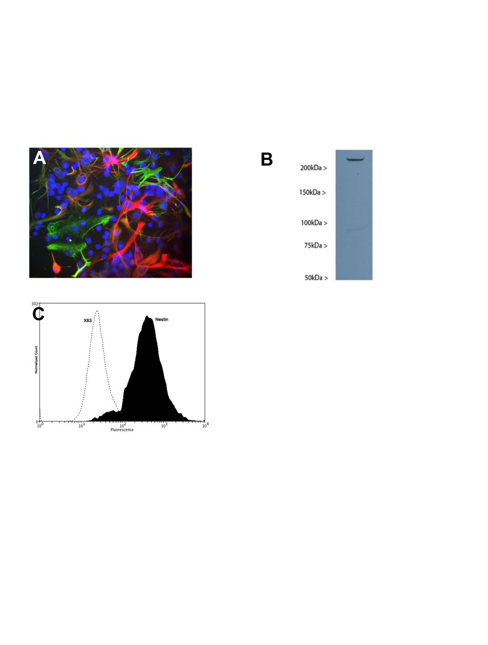
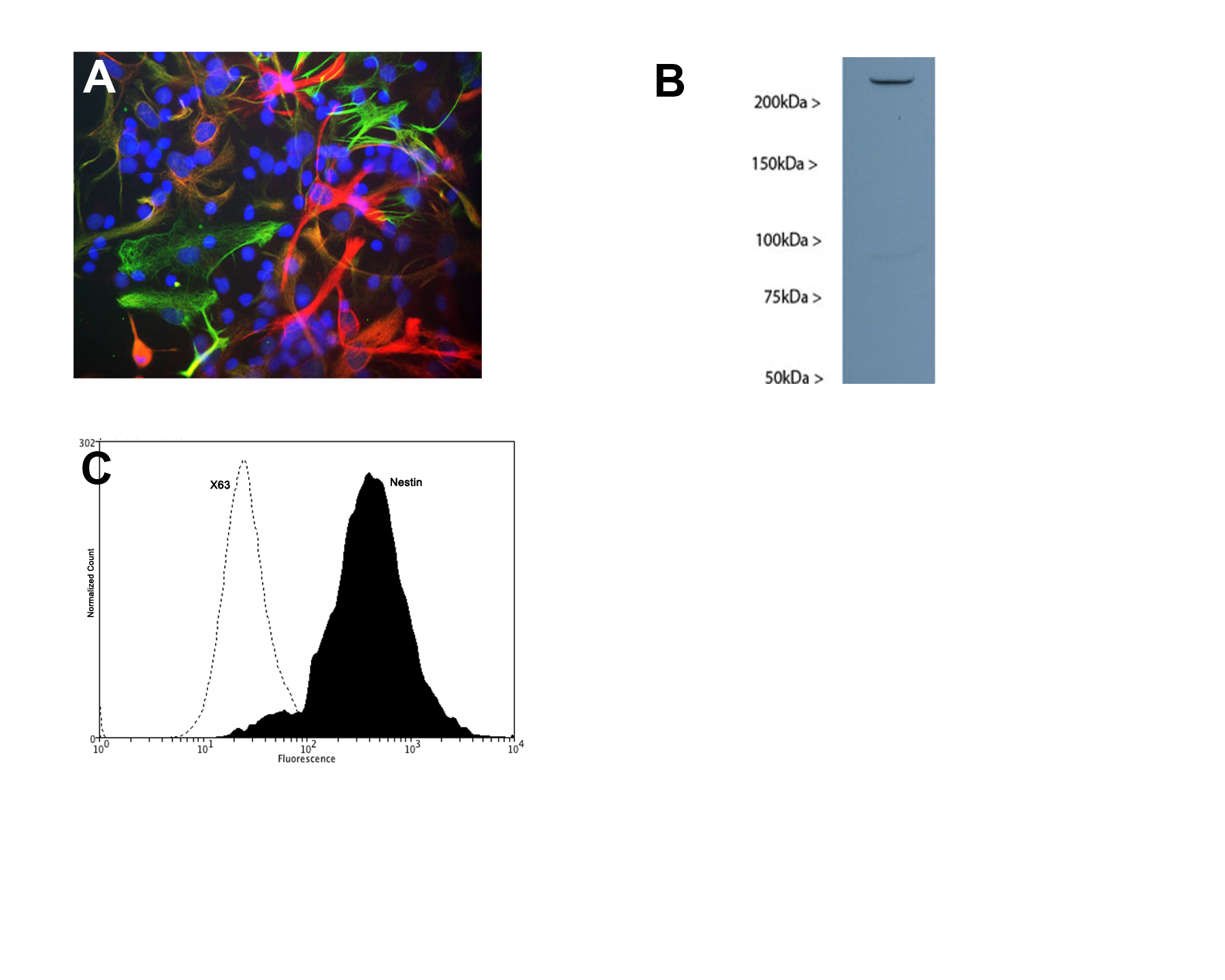
A: Mixed cultures of neonatal rat neurons and glia stained with Mouse monoclonal antibody to Nestin [4D11] M-1385-100 (red), Chicken polyclonal antibody to vimentin C-1409-50 (green) and DNA (DAPI stain, blue). Astrocytes and neuronal stem cells stain strongly and specifically in a clearly filamentous fashion with the Nestin antibody. The filamentous staining pattern is as expected as both Nestin and Vimentin are components of 10nm filaments. Note that some cells contain Nestin, but do not stain strongly for Vimentin and so appear red. Others contain Vimentin and not Nestin and so appear green- these are likely to be fibroblastic or endothelial cells. Some cells express both proteins and so appear yellowish. The presence of Nestin indicates that the cells are developing astrocytes, neuroblasts or undifferentiated neural stem cells. B: Western blot in of developing rat brain (P18) homogenate probed with mouse monoclonal antibody to Nestin M-1385-100. A single strong band running at ~240 kDa is seen. C: Analysis of nesting expression in human neuroblastoma SH-SY5Y by Flow Cytometry. Fixing and Permeabilization of cells: Absolute methanol (10 minutes in ice) and 0.1% Tween-20 in PBS, Blocking: 1% BSA, Primary antibody: Mouse Monoclonal antibody to Nestin (cat # M-1385-100, 2μg per ~10^6 cells) for 30 minutes at room temperature, Secondary antibody: Goat anti-mouse PE labeled secondary antibody (1:100 fold dilution) with incubation for 20 minutes in dark at room temperature. Non-specific Control IgG, clone X63 (cat # M-1249-200) was used as negative control under same conditions (black dashed). Flow cytometry data and results were generated using Orflo MoxiflowTM instrument and protocols.
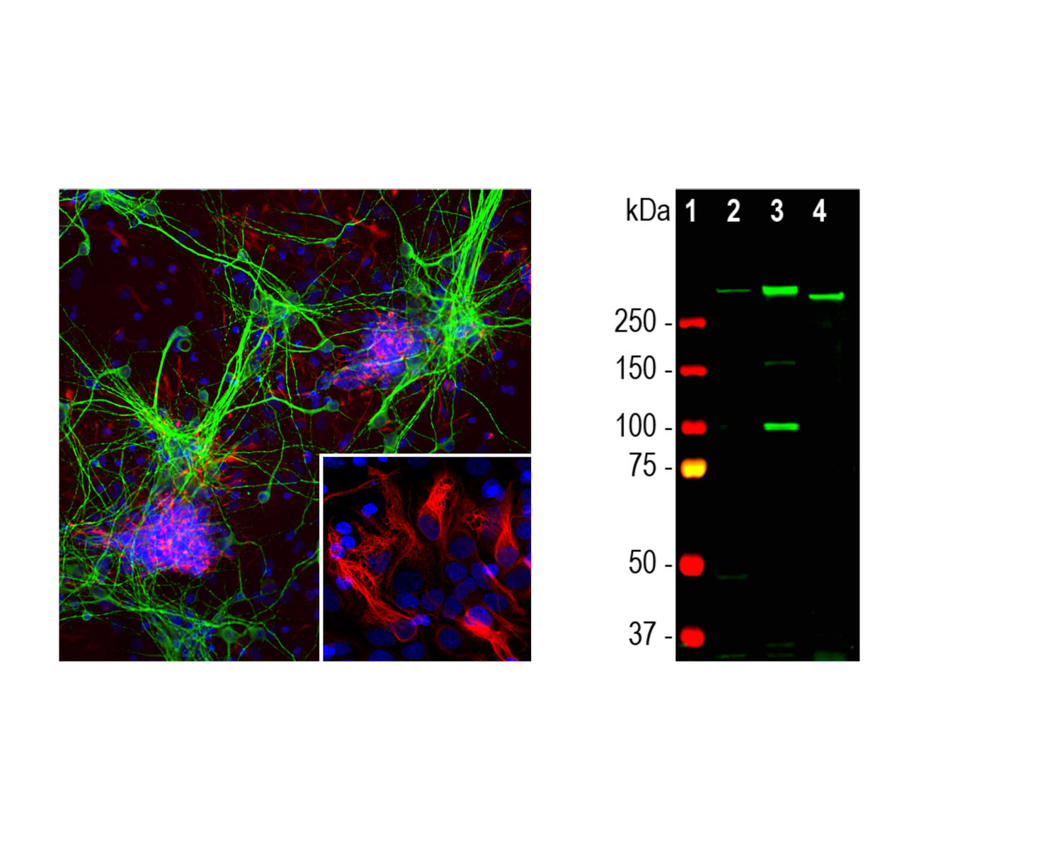
Left: Detection of nestin expression in cortical neuron-glial cell culture from E20 rat by Immunocytochemistry. Cells were stained with mouse anti-nestin antibody (1:500, red) and with chicken antibody to MAP2 (C-1382-50, 1:5,000, green). Blue: DAPI nuclear dye. The nestin antibody labels developing astrocytes and neuronal stem cells in a clearly filamentous fashion, while the MAP2 antibody stains dendrites and perikarya of mature neurons. Right: Western blot analysis of tissue and cell lysates with mouse antibody to nestin (1:500, green). [1] protein standard, [2] embryonic E18 rat brain, [3] C6 rat glioma cells, and [4] SH-SY5Y human neuroblastoma cells.
Specific ReferencesSchomann T et al. (2020) Multimodal imaging of hair follicle bulge-derived stem cells in a mouse model of traumatic brain injury. Cell Tissue Res. [Epub ahead of print]. Application: IHC/IF; Species: Mouse.
Schomann T et al. (2017) Neuronal differentiation of hair-follicle-bulge-derived stem cells co-cultured with mouse cochlear modiolus explants. PLos One. 12(10):e0187183. Application: ICC/IF; Species: Mouse, Hair follicle bulge-derived neural crest-derived stem cells (HFBSCs).
Gho CG et al. (2015) Isolation, expansion and neural differentiation of stem cells from human plucked hair- a further step towards autologous nerve recovery.Cytotechnology In press. Application: IF; Species: Human, Hair follicle bulge-derived neural crest-derived stem cells (HFBSCs), Keywords: Hair follicle stem cell, Regeneration, Neural crest, Neuron, Glia, Cryopreservation
 1800 605-5127
 +61 (0)8 8352 7711