Product DescriptiongoogleRabbit anti-Laminin-111 Polyclonal Antibody (Unconjugated), suitable for WB, IHC-Frozen.
Alternative NamesLaminin 1, Laminin _1_1_1.
Application(s)IHC-Frozen, WB
Antibody HostRabbit
Antibody TypePolyclonal
SpecificityMouse,reacts with Mouse and Rat, other species not tested. This antibody recognizes laminin isotypes alpha-1 (440 kDa), beta-1 (220 kDa) and gamma-1 (220 kDa). It also binds laminin binding protein at 120 kDa which always co-expresses with laminin.
Species ReactivityMouse, Rat
Immunogen DescriptionLaminin-111 isolated from mouse EHS cells
Product DescriptionRabbit anti-Laminin-111 Polyclonal Antibody (Unconjugated), suitable for WB, IHC-Frozen.
Application(s)IHC-Frozen, WB
Application DetailsWestern blotting (1:1,000-1:5,000) and Immunohistochemistry (1:1,000-1:5,000). Biosensis recommends optimal dilutions/concentrations should be determined by the end user.
TargetLaminin-111
SpecificityMouse,reacts with Mouse and Rat, other species not tested. This antibody recognizes laminin isotypes alpha-1 (440 kDa), beta-1 (220 kDa) and gamma-1 (220 kDa). It also binds laminin binding protein at 120 kDa which always co-expresses with laminin.
Target Host SpeciesMouse
Species ReactivityMouse, Rat
Antibody HostRabbit
Antibody TypePolyclonal
Antibody IsotypeIgG
ConjugateUnconjugated
Immunogen DescriptionLaminin-111 isolated from mouse EHS cells
Purity DescriptionAffinity purified
FormatLyophilized from PBS buffer pH 7.2-7.6 with 0.1% trehalose, and sodium azide
Reconstitution InstructionsSpin vial briefly before opening. Reconstitute with 100 µL sterile-filtered, ultrapure water. Centrifuge to remove any insoluble material.
Storage InstructionsStore lyophilized antibody at 2-8°C. After reconstitution divide into aliquots and store at -20°C for long-term storage. Store at 2-8°C short-term (up to 4 weeks) with an appropriate antibacterial agent. Avoid repetitive freeze/thaw cycles.
Batch NumberPlease see item label.
Expiration Date12 months after date of receipt (unopened vial).
Scientific BackgroundBinding to cells via a high affinity receptor, laminin is thought to mediate the attachment, migration and organisation of cells into tissues during embryonic development by interacting with other extracellular matrix components. Ref: uniprot.org
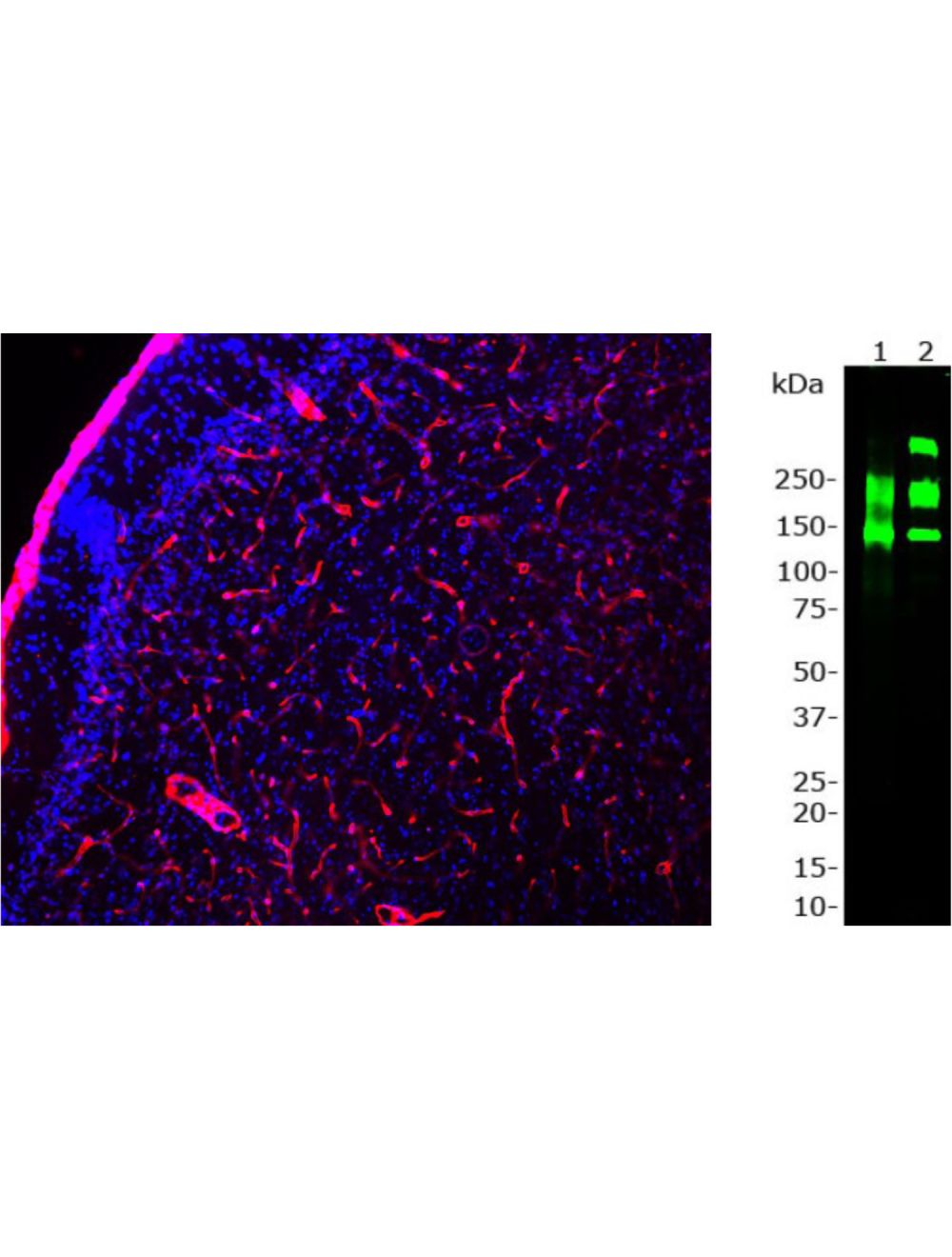
Left: Staining of laminin in mouse section of cortex by Immunohistochemistry. Rabbit antibody to Laminin (red) reveals strong staining in the basement membranes of blood vessels. Blue: DAPI nuclear stain. Right: Western blot analysis of rat heart cell lysate (Lane 1) and 0.2 ug of purified laminin-111 protein from mouse EHS sarcoma (Lane 2). This antibody recognizes 3 laminin isotypes: alpha-1 (440 kDa), beta-1 (220 kD) and gamma-1 (220 kDa). Also recognized is a laminin-binding protein at 120 kDa in both rat heart lysate and purified laminin protein. Since this protein always co-expresses with laminin this crossreactivity is irrelevant.
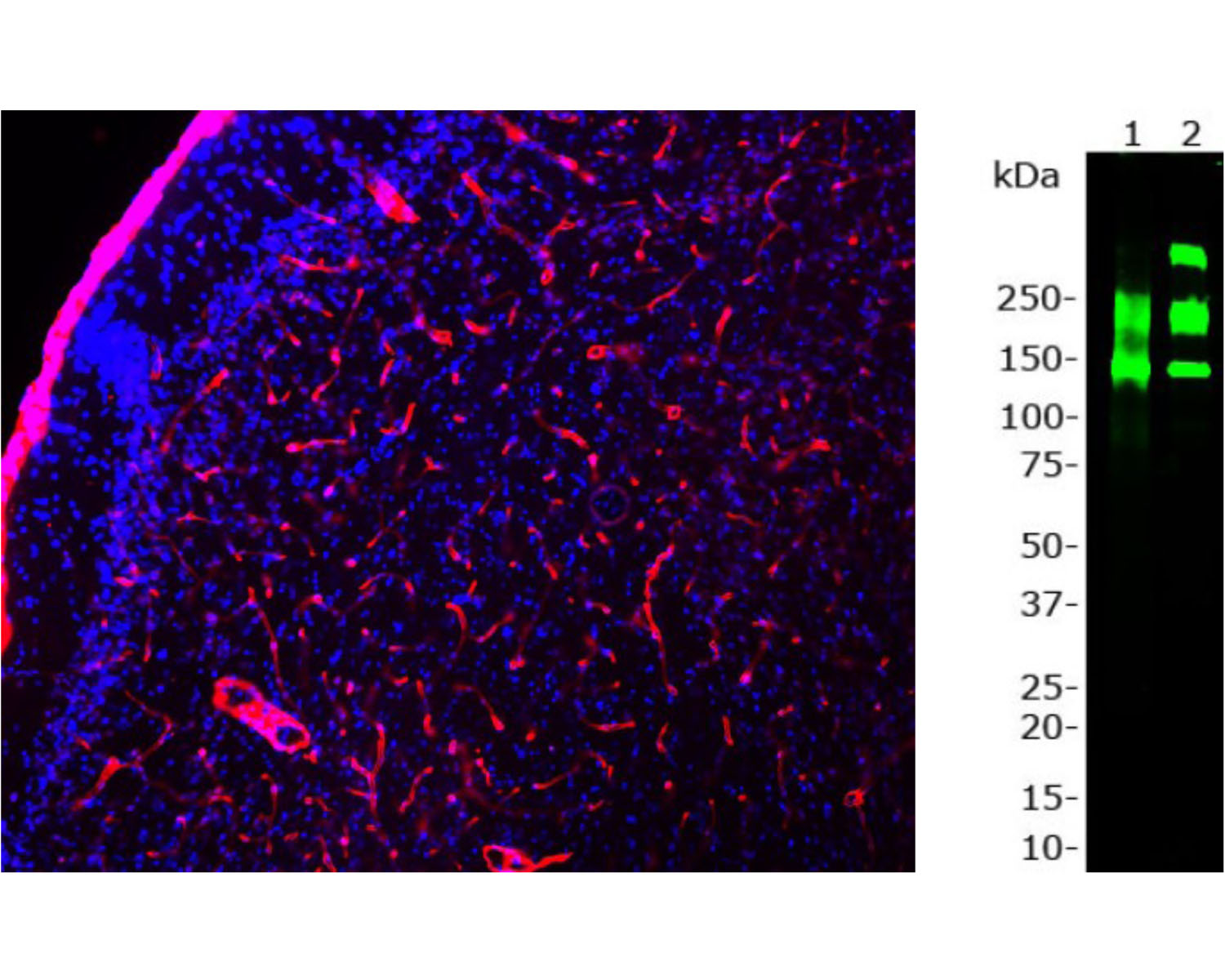
Left: Analysis of rat brain stem section stained with rabbit anti-laminin (red, 1:1,000), and chicken anti-myelin basic protein (MBP, C-1383-50, green, 1:5,000) by Immunohistochemistry. Blue: DAPI nuclear stain. IHC Method: Following transcardial perfusion of rat with 4% paraformaldehyde, brain was post fixed for 24 hours, cut to 45 um, and free-floating sections were stained.The laminin antibody is an excellent marker of basement membranes surrounding blood vessels, while the MBP antibody stains the myelin sheathes around axons. Right: Western blot analysis of laminin expression (green, 1:5,000). [2] rat brain, [3] rat spinal cord, and [4] cow spinal cord. The strong band above the 250 kDa mark corresponds to full-length laminin protein. Smaller proteolytic fragments of laminin are also detected with this antibody.
 1800 605-5127
 +61 (0)8 8352 7711