Product DescriptionRabbit anti-Neuron specific enolase (NSE) Polyclonal Antibody (Unconjugated), suitable for WB, ICC.
Application(s)ICC, WB
Application DetailsWestern Blotting (WB) and Immunocytochemistry (ICC). A dilution of 1:1,000 - 1:2,000 is recommended for WB. A dilution of 1:500 is recommended for ICC. Biosensis recommends optimal dilutions/concentrations should be determined by the end user.
TargetNeuron specific enolase (NSE)
SpecificitySpecifically recognizes ~47 kDa NSE protein in WB. Human and Rat. Predicted to react with other mammals due to sequence homology.
Target Host SpeciesHuman
Species ReactivityHuman, Mouse, Rat
Antibody HostRabbit
Antibody TypePolyclonal
Antibody IsotypeMixed
ConjugateUnconjugated
Immunogen DescriptionRecombinant human Neuron Specific Enolase (NSE) expressed in and purified from E.coli
Purity DescriptionWhole serum
FormatLyophilized with sodium azide.
Reconstitution InstructionsSpin vial briefly before opening. Reconstitute with 50 µL sterile-filtered, ultrapure water. Centrifuge to remove any insoluble material.
Storage InstructionsAfter reconstitution of lyophilized antibody, aliquot and store at -20°C for a higher stability. Avoid freeze-thaw cycles.
Batch NumberPlease see item label.
Expiration Date12 months after date of receipt (unopened vial).
Scientific BackgroundEnolase is a metalloenzyme that catayzes the reaction between 2-phospho-D-glycerate and phosphoenolpyruvate during glycolysis. Mammalian enolase is composed of 3 subunits; alpha, beta and gamma (Neuron-specific enolase). These subunits can form homodimers or heterodimers. The alpha/gamma heterodimer and the gamma/gamma homodimer are found primarily in neurons.
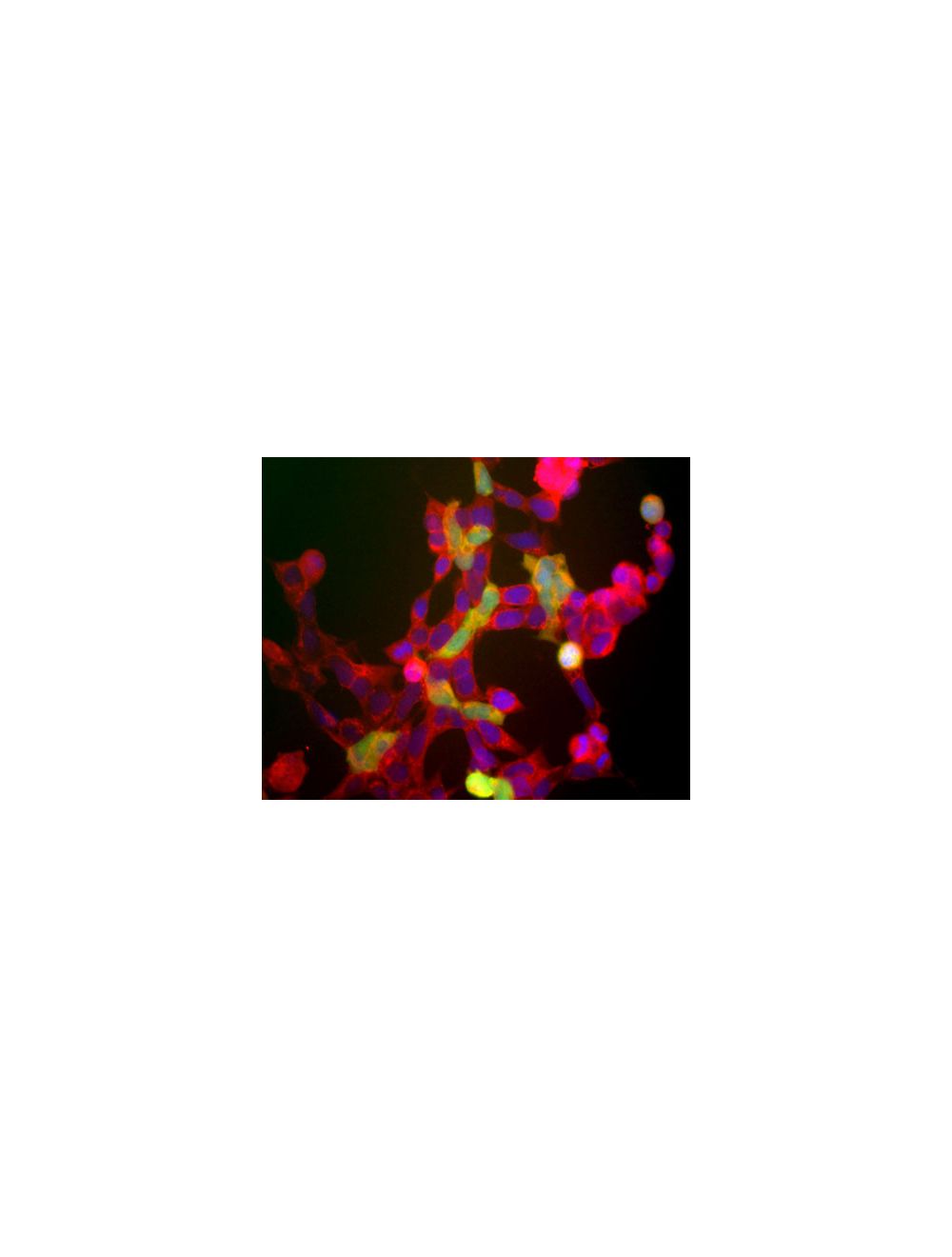
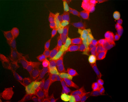
Image shows the human embryonic kidney cells line 293 (Hek293). The red channel shows staining with Rabbit polyclonal antibody to Neuron specific enolase R-1396-50, which recognizes all of these 293 cells. The green channels shows staining for another neuronal marker with Mouse monoclonal antibody to ubiquitin C-terminal hydrolase 1 (UCHL1), M-1407-100. This neuronal gene is apparently activated in a cell density dependent fashion and at this stage only a few cells express this protein. However all cells that express NSE also express UCHL1.
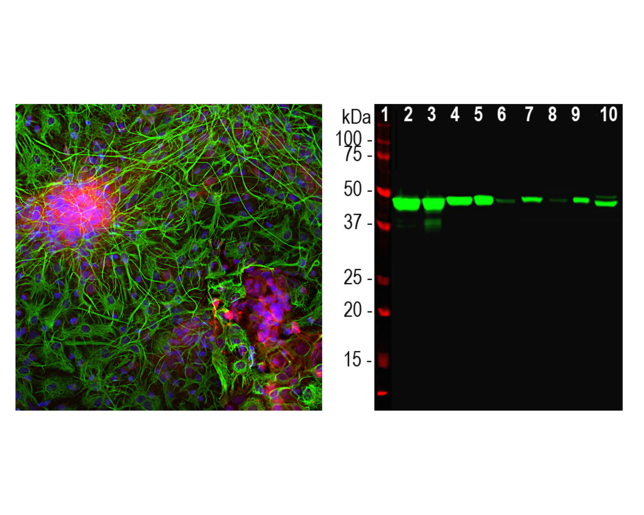
Left: Immunofluorescent analysis of mixed cortical neuron-glial cell culture from E20 rat by Immunocytochemistry. Cells were stained with rabbit antibody to NSE (red, 1:500), and co-stained with chicken antibody to GFAP (C-1373-50, green, 1:5,000). Blue: Hoechst nuclear dye. The NSE antibody labels protein expressed in neuronal cells, while the GFAP antibody stains intermediate filaments in astrocytes. Right: Western blot analysis of tissue and cell lysates using rabbit antibody to NSE (green, 1:5,000). [1] protein standard, [2] rat brain, [3] rat spinal cord, [4] mouse brain, [5] mouse spinal cord, [6] NIH-3T3, [7] HEK293, [8] HeLa, [9] SH-SY5Y, and [10] C6. A single band at about 47 kDa corresponds to the NSE protein, seen only in extracts containing neurons or neuronal lineage cells.
 1800 605-5127
 +61 (0)8 8352 7711