Product DescriptionRabbit anti-Ki-67 Polyclonal Antibody (Unconjugated), suitable for WB, ICC.
Application(s)ICC, WB
Application DetailsWestern blotting (1:2,000-1:10,000) and Immunocytochemistry (1:1,000-1:5,000). Biosensis recommends optimal dilutions/concentrations should be determined by the end user.
TargetKi-67
SpecificityHuman,reacts with Human only. Does not react with Mouse or Rat.
Target Host SpeciesHuman
Species ReactivityHuman
Antibody HostRabbit
Antibody TypePolyclonal
Antibody IsotypeIgG
ConjugateUnconjugated
Immunogen DescriptionRecombinant human Ki-67 protein (amino acids 1,111-1,490) expressed in and purified from E. coli.
Purity DescriptionWhole serum
FormatLyophilized with sodium azide.
Reconstitution InstructionsSpin vial briefly before opening. Reconstitute with 100 µL sterile-filtered, ultrapure water. Centrifuge to remove any insoluble material.
Storage InstructionsStore lyophilized antibody at 2-8°C. After reconstitution divide into aliquots and store at -20°C for long-term storage. Store at 2-8°C short-term (up to 4 weeks) with an appropriate antibacterial agent. Avoid repetitive freeze/thaw cycles.
Batch NumberPlease see item label.
Expiration Date12 months after date of receipt (unopened vial).
Scientific BackgroundRequired to maintain individual mitotic chromosomes dispersed in the cytoplasm following nuclear envelope disassembly (PubMed:27362226). Associates with the surface of the mitotic chromosome, the perichromosomal layer, and covers a substantial fraction of the chromosome surface (PubMed:27362226). Prevents chromosomes from collapsing into a single chromatin mass by forming a steric and electrostatic charge barrier: the protein has a high net electrical charge and acts as a surfactant, dispersing chromosomes and enabling independent chromosome motility (PubMed:27362226). Binds DNA, with a preference for supercoiled DNA and AT-rich DNA (PubMed:10878551). Does not contribute to the internal structure of mitotic chromosomes (By similarity). May play a role in chromatin organization (PubMed:24867636). It is however unclear whether it plays a direct role in chromatin organization or whether it is an indirect consequence of its function in maintaining mitotic chromosomes dispersed (Probable). Ref: uniprot.org
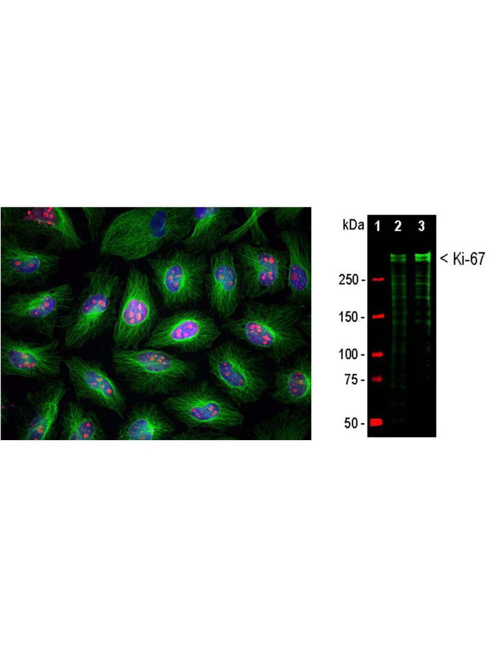
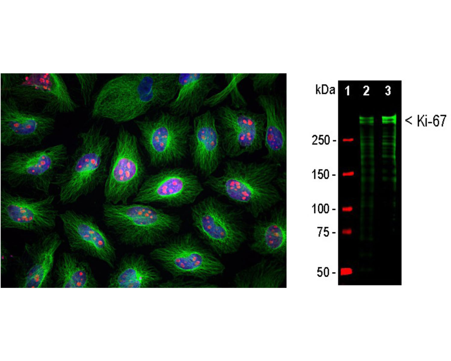
Left: Immunofluorescence analysis of HeLa cells costained with rabbit antibody to Ki-67 (1:5,000, red) and a mouse antibody to beta-tubulin. Ki-67 is predominantly localized in nucleoli of cells in interphase, while in mitotic cells Ki-67 is associated with condensed chromosomes. Ki-67 is not expressed in cells in quiescent G0 state. Blue: DAPI staining of nuclear DNA. Right: Western blots analysis of equal amounts of HeLa (Lane 2) and HEK293 (Lane 3) cell lysates using rabbit antibody to Ki-67 (1:10,000). Double bands above 250 kDa correspond to two major Ki-67 isoforms of molecular weight ~345 and 395 kDa. Smaller fragments of these isoforms are also detected on the blot.
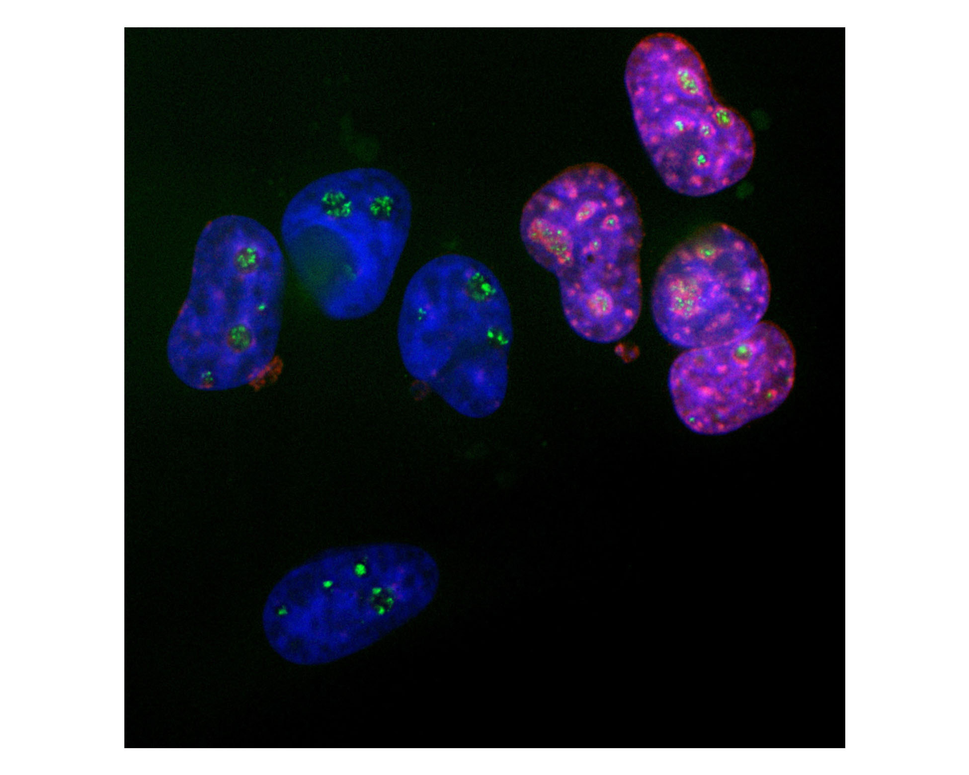
Analysis of Ki67 expression in cultured HeLa cells with rabbit anti-Ki67 antibody (red, 1:5,000), and mouse anti-fibrillarin (M-1372-250, green, 1:2,000) by Immunocytochemistry. Blue: DAPI nuclear stain. Ki67 protein accumulates in and around the nucleoli of interphase cells such as those on the right, and the nucleoli are revealed by the fibrillarin antibody. In contrast, cells in the quiescent G0 state such as those on the left are Ki67 negative, but fibrillarin-positive.
 1800 605-5127
 +61 (0)8 8352 7711