1800 605-5127
 +61 (0)8 8352 7711
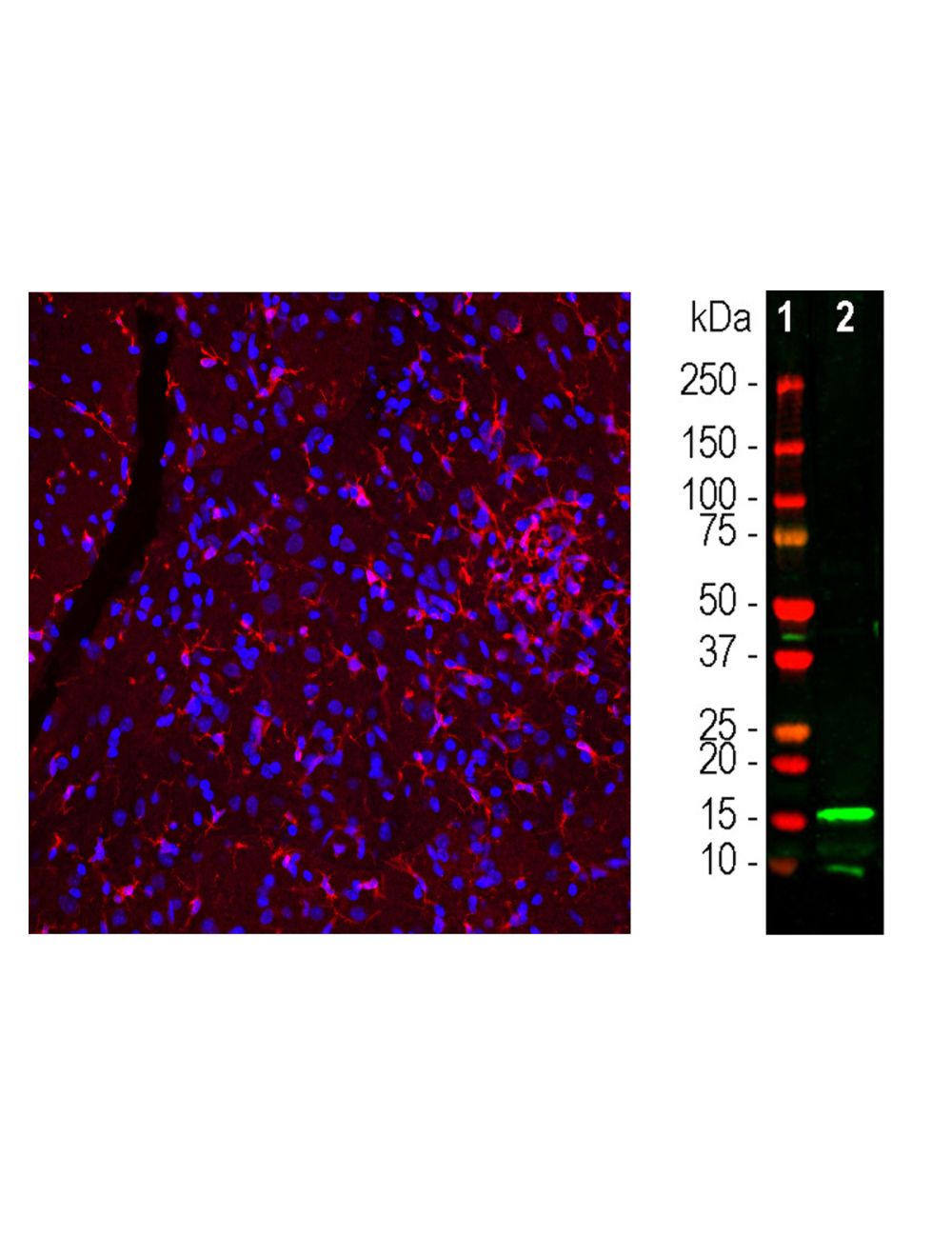

Ionized Calcium Binding Adapter Molecule 1 (IBA1), Chicken Polyclonal Antibody
As low as
US$317.00
Only %1 left
Catalog Number
C-2118
- Product Name Ionized Calcium Binding Adapter Molecule 1 (IBA1), Chicken Polyclonal Antibody
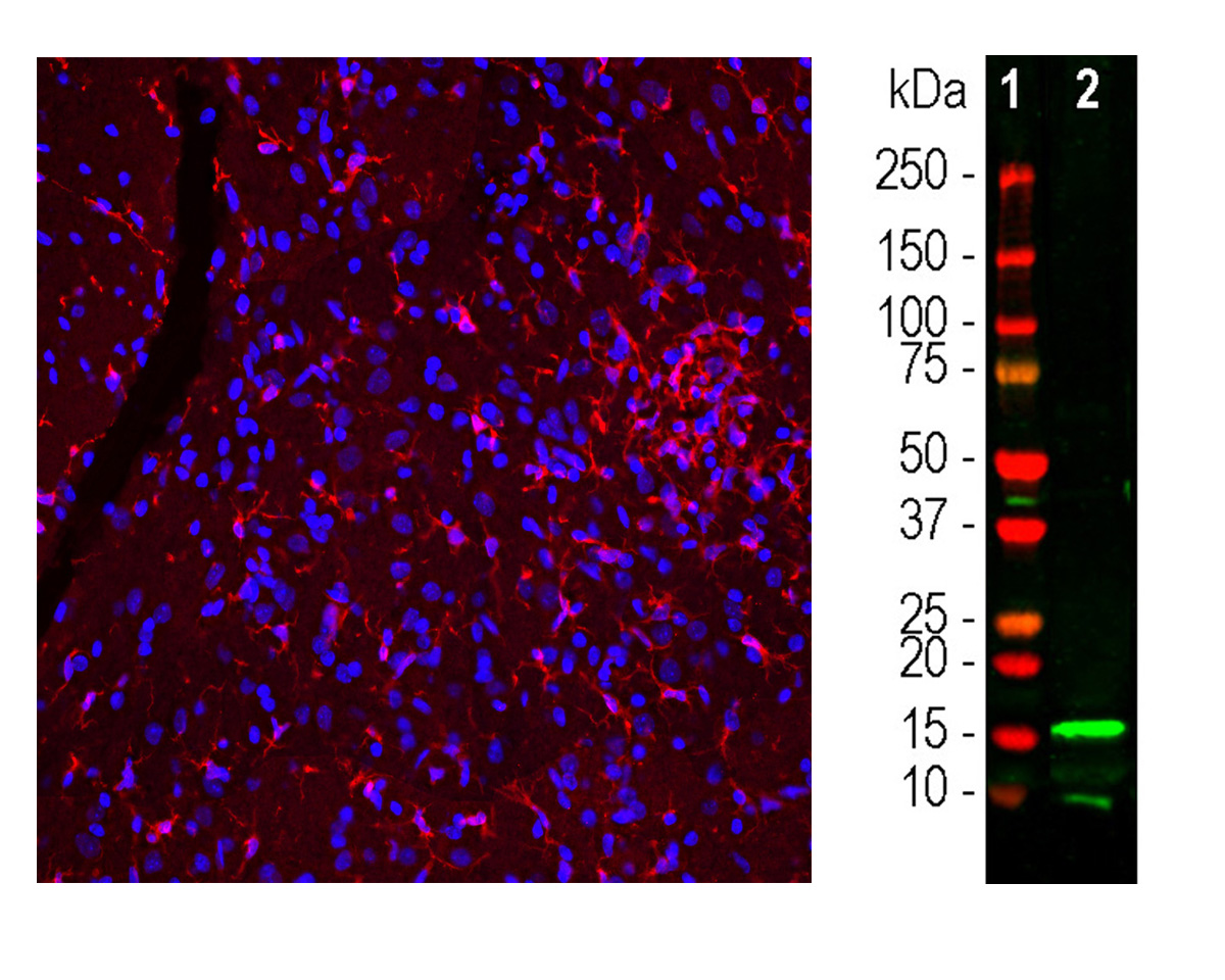
- Product Description Chicken anti-Ionized Calcium Binding Adapter Molecule 1 (IBA1), Polyclonal Antibody (Unconjugated), suitable for WB, IHC-Frozen, ICC
- Alternative Names IBA-1; AIF1; AIF-1 G1; Allograft inflammatory factor 1; Ionized calcium binding adapter molecule 1; Protein G1
- Application(s) ICC, IHC-Frozen, WB
- Antibody Host Chicken
- Antibody Type Polyclonal
- Specificity Human. Species cross-reactivity includes rat and mouse.
- Species Reactivity Human, Mouse, Rat
- Immunogen Description C-terminal peptide of human IBA1. The antibody has been made against the C-terminal peptide of human IBA1 coupled to keyhole limpet hemocyanin (KLH).
- Conjugate Unconjugated
- Purity Description IgY Fraction
- Regulatory Status For research use only.
Product Info
- Product Description Chicken anti-Ionized Calcium Binding Adapter Molecule 1 (IBA1), Polyclonal Antibody (Unconjugated), suitable for WB, IHC-Frozen, ICC
-
Related Products
Ionized calcium-binding adapter molecule 1 (IBA1), Rabbit Polyclonal Antibody
- Application(s) ICC, IHC-Frozen, WB
- Application Details Western Blotting (WB), Immunocytochemistry (ICC), Immunohistochemistry (IHC). A dilution of 1:5,000 - 1:10,000 is recommended for WB. A dilution of 1:100 - 1:500 is recommended for IC and IH. Biosensis recommends optimal dilutions/concentrations should be determined by the end user.
- Target Ionized calcium binding adapter molecule 1 (IBA1)
- Specificity Human. Species cross-reactivity includes rat and mouse.
- Target Host Species Human
- Species Reactivity Human, Mouse, Rat
- Antibody Host Chicken
- Antibody Type Polyclonal
- Antibody Isotype IgY
- Conjugate Unconjugated
- Immunogen Description C-terminal peptide of human IBA1. The antibody has been made against the C-terminal peptide of human IBA1 coupled to keyhole limpet hemocyanin (KLH).
- Purity Description IgY Fraction
- Format Lyophilized IgY preparation, with sodium azide.
- Reconstitution Instructions Spin vial briefly before opening. Reconstitute with 50 µL sterile-filtered, ultrapure water. Centrifuge to remove any insoluble material.
- Storage Instructions After reconstitution of lyophilized antibody, aliquot and store at -20°C for a higher stability. Avoid freeze-thaw cycles. Store at 4°C for up to one month for short term storage and frequent use.
- Batch Number Please see item label.
- Expiration Date 12 months after date of receipt (unopened vial).
- Alternative Names IBA-1; AIF1; AIF-1 G1; Allograft inflammatory factor 1; Ionized calcium binding adapter molecule 1; Protein G1
- Uniprot Number P55008
- Uniprot Number/Name P55008 (AIF1_HUMAN)
- Scientific Background Actin-binding protein that enhances membrane ruffling and RAC activation. Enhances the actin-bundling activity of LCP1. Binds calcium. Plays a role in RAC signaling and in phagocytosis. May play a role in macrophage activation and function. Promotes the proliferation of vascular smooth muscle cells and of T-lymphocytes. Enhances lymphocyte migration. Plays a role in vascular inflammation. (Ref: uniprot.org)
- Shipping Temperature 25°C (ambient)
- UNSPSC CODE 41116161
- Regulatory Status For research use only.