Product DescriptionMouse anti-Glial Fibrillary Acidic Protein (GFAP) Monoclonal Antibody (Unconjugated), suitable for WB, IHC-Frozen.
Application(s)IHC-Frozen, WB
Application DetailsWestern Blot (1:1,000-1:2,000): tested on rat, mouse brain and spinal cord, human recombinant protein, pig brain. Immunohistochemistry (1:500-1:1,000): tested on rat cerebellum section. Other applications not yet tested. Biosensis recommends optimal dilutions/concentrations should be determined by the end user.
TargetGlial Fibrillary Acidic Protein (GFAP)
SpecificityThis antibody is specific for GFAP as demonstrated by western blotting and immunohistochemistry.
Target Host SpeciesPig
Species ReactivityBovine, Human, Mouse, Pig, Rat
Antibody HostMouse
Antibody TypeMonoclonal
Antibody IsotypeIgG1
Clone Name2A5
ConjugateUnconjugated
Immunogen DescriptionGFAP isolated biochemically from pig spinal cord was used as the immunogen.
Purity DescriptionProtein G purified
FormatLyophilized from PBS buffer pH 7.2-7.6 with 0.1% trehalose, and sodium azide
Reconstitution InstructionsSpin vial briefly before opening. Reconstitute with 100 µL sterile-filtered, ultrapure water to achieve a 1 mg/mL concentration. Centrifuge to remove any insoluble material.
Storage InstructionsStore lyophilized antibody at 2-8°C. After reconstitution divide into aliquots and store at -20°C for long-term storage. Store at 2-8°C short-term (up to 4 weeks) with an appropriate antibacterial agent. Avoid repetitive freeze/thaw cycles.
Batch NumberPlease see item label.
Expiration Date12 months after date of receipt (unopened vial).
Alternative NamesGlial fibrillary acidic protein; GFAP
Scientific BackgroundGFAP is a 50 kDa intra-cytoplasmic filamentous protein of the cytoskeleton in astrocytes. During the development of the central nervous system, it is a cell-specific marker that distinguishes astrocytes from other glial cells. GFAP immunoreactivity has been shown in immature oligodendrocytes, epiglottic cartilage, pituicytes, papillary meningiomas, myoepithelial cells of the breast and in non-CNS: Schwann cells, salivary gland neoplasms, enteric glia cells, and metastasizing renal carcinomas.
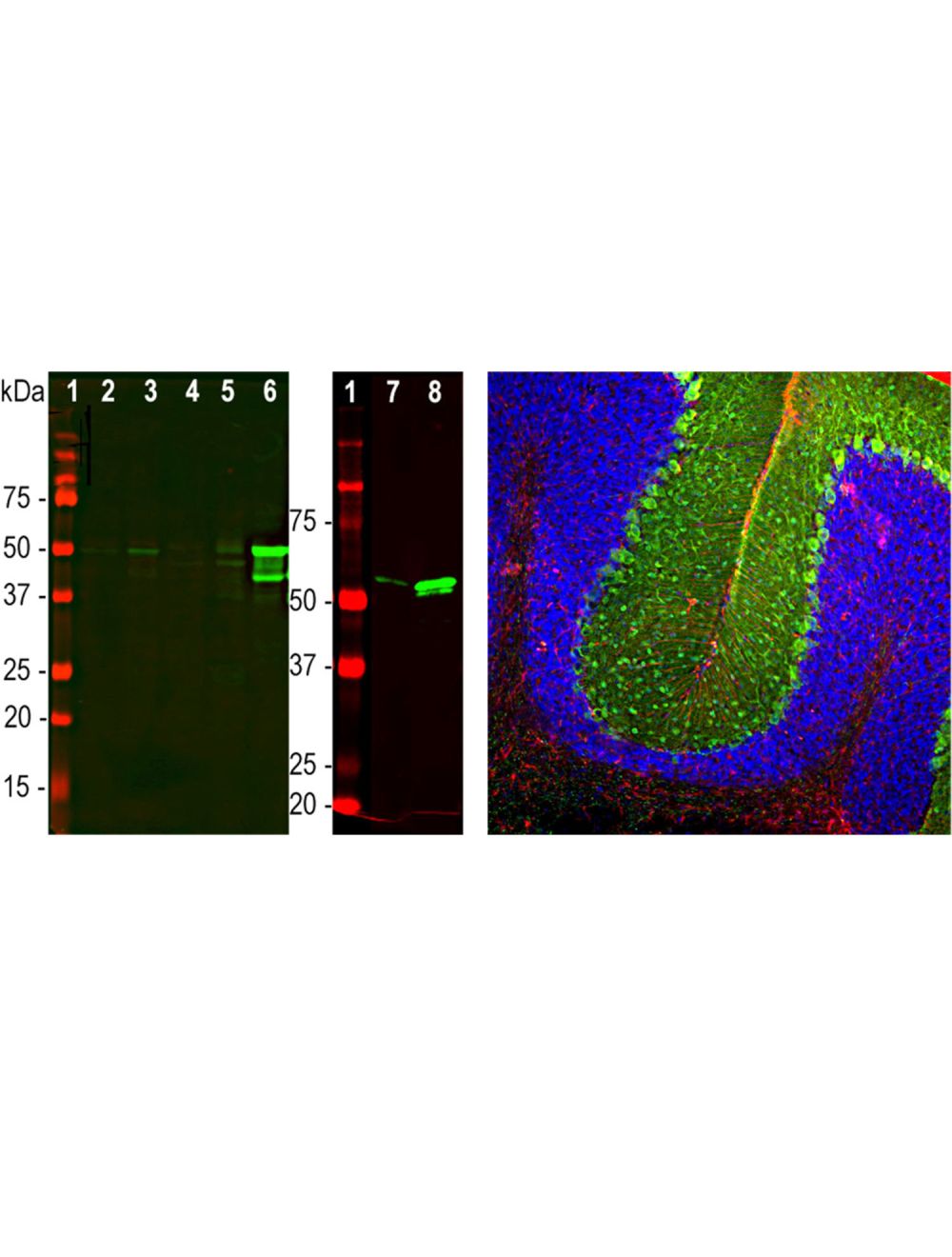
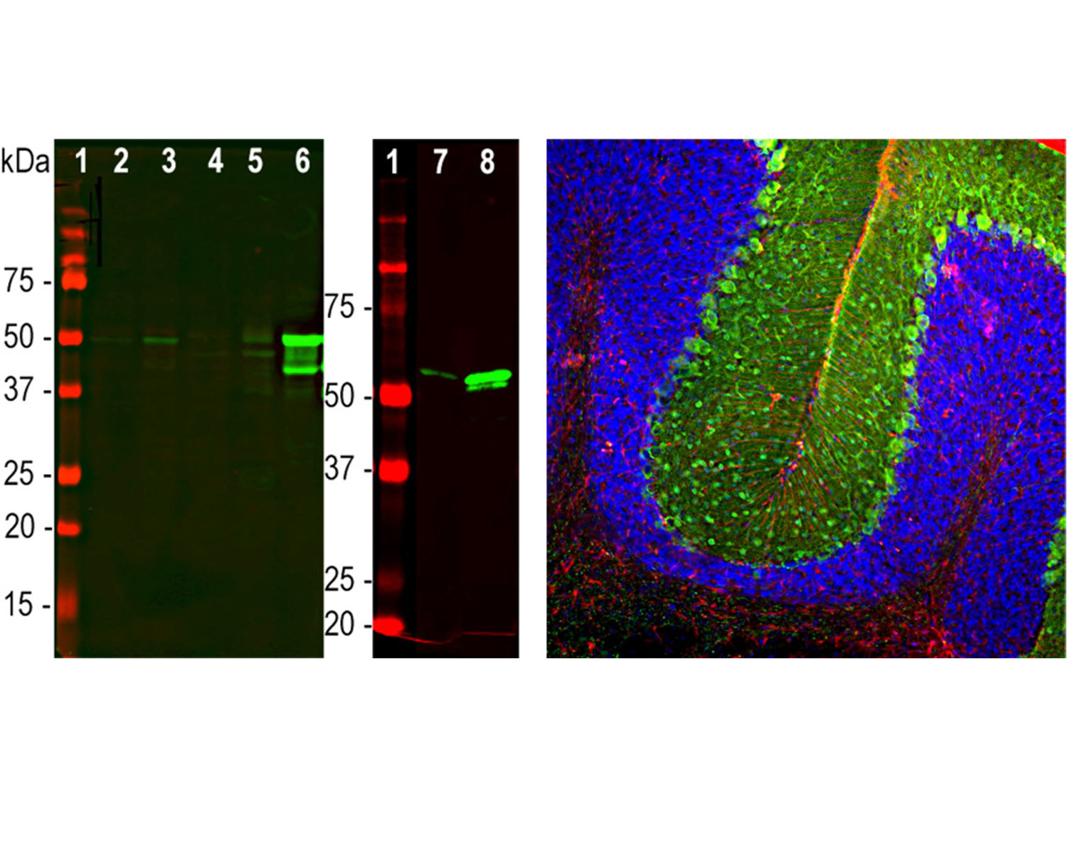
Left: Western blot analysis of GFAP expression in tissue homogenates. [1] protein standard, [2] rat brain, [3] rat spinal cord, [4] mouse brain, [5] mouse spinal cord, [6] pig brain, [7] rat recombinant GFAP, [8] human recombinant GFAP. Bands around 50 kDa correspond to alternative transcripts and proteolytic products of GFAP. Primary antibody was diluted 1:2,000. Note that this antibody has significantly stronger reactivity with pig and human GFAP as compared to rodent, suggesting that it binds to an epitope which is not totally conserved across mammalian sequences. Right: Analysis of GFAP expression in an adult rat cerebellum section by Immunohistochemistry. Primary antibodies: mouse anti-GFAP (red, 1:500), chicken anti-Parvalbumin (C-1814-50, green, 1:2,000). Blue: DAPI nuclear stain. IHC Method: Following transcardial perfusion of rat with 4% paraformaldehyde, brain was post-fixed for 24 hours, and free-floating 45 uM sections were stained. The GFAP antibody stains the processes of Bergmann glia and astrocytes. The Parvalbumin antibody labels perikarya and dendrites of Purkinje cells and interneurons in the molecular layer of the cerebellum. The staining on rodent tissues is specific but not as robust as on human material.
 1800 605-5127
 +61 (0)8 8352 7711