Product NameCalretinin-binding protein (CR), Mouse Monoclonal Antibody
Product DescriptiongoogleMouse anti-Calretinin-binding protein (CR) Monoclonal Antibody (Unconjugated), suitable for WB, IHC-Frozen.
Alternative NamesCR, 29 kDa calbindin
Application(s)IHC-Frozen, WB
Antibody HostMouse
Antibody TypeMonoclonal
SpecificityHuman, reacts with Human, Cow, Rat, Mouse. Antibody is specific for calbindin and does not recognize closely related proteins parvalbumin, calretinin and secretagogin as determined by Western Blotting.
Species ReactivityBovine, Human, Mouse, Rat
Immunogen DescriptionFull-length recombinant human protein
Product DescriptionMouse anti-Calretinin-binding protein (CR) Monoclonal Antibody (Unconjugated), suitable for WB, IHC-Frozen.
Application(s)IHC-Frozen, WB
Application DetailsWestern blotting (1:1,000-1:5,000) and Immunohistochemistry (1:1,000). Biosensis recommends optimal dilutions/concentrations should be determined by the end user.
TargetCalretinin-binding protein (CR)
SpecificityHuman, reacts with Human, Cow, Rat, Mouse. Antibody is specific for calbindin and does not recognize closely related proteins parvalbumin, calretinin and secretagogin as determined by Western Blotting.
Target Host SpeciesHuman
Species ReactivityBovine, Human, Mouse, Rat
Antibody HostMouse
Antibody TypeMonoclonal
Antibody IsotypeIgG1
Clone Name3G9
ConjugateUnconjugated
Immunogen DescriptionFull-length recombinant human protein
Purity DescriptionProtein G purified
FormatLyophilized from PBS buffer pH 7.2-7.6 with 0.1% trehalose, and sodium azide
Reconstitution InstructionsSpin vial briefly before opening. Reconstitute with 100 µL sterile-filtered, ultrapure water to achieve a 1 mg/mL concentration. Centrifuge to remove any insoluble material.
Storage InstructionsStore lyophilized antibody at 2-8°C. After reconstitution divide into aliquots and store at -20°C for long-term storage. Store at 2-8°C short-term (up to 4 weeks) with an appropriate antibacterial agent. Avoid repetitive freeze/thaw cycles.
Batch NumberPlease see item label.
Expiration Date12 months after date of receipt (unopened vial).
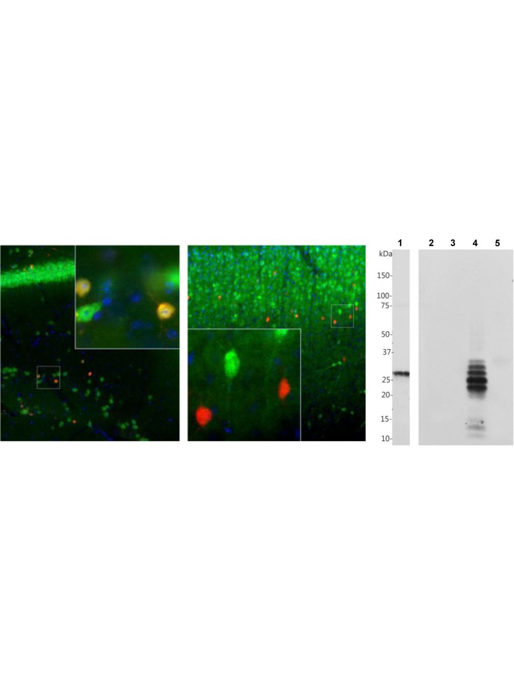
Left and Middle: Detection of calretinin immunoreactivity (red) in adult mouse brain hippocampal sections (Left) and adult rat cortical sections (Middle) by Immunohistochemistry. The calretinin antibody (1:1,000) stains a small number of interneurons in the stratum radiatum of CA1 region (Left), while Fox/NeuN (R-3770-100, green) is expressed in most neurons in the brain. As a result, co-labelled neurons that stain positive for calretinin appear yellow. In rat cortex (Right), calretinin (red) is expressed in a small population of interneurons concentrated in Layer 4 area, while calbindin (C-1798-50, green) is expressed in cells concentrated in Layer 2/3. Because each antibody specifically labels a different population of cells exclusively, the cells are either stained green or red in the cortex. Blue: DAPI nuclear stain. Insets are high-magnification images of the boxed area in each picture. IHC method: 45 um sections, tissue fixed by transcardial perfusion with 4% paraformaldehyde. Right: Western blot analysis of calretinin. The antibody binds strongly and cleanly to a calretinin band at ~32 kDa in cow cerebellum homogenate (Lane 1). Specificity for calretinin and absecnce of cross-reactivity with other related calcium-binding proteins is shown by probing recombinant proteins: secretagogin (Lane 2), parvalbumin (Lane 3), calretinin (Lane 4), calbindin (Lane 5).
Rat cerebellum stained with mouse anti-calretinin (green, 1:500) and chicken anti-calbindin (C-1798-50, red, 1:2,000) by Immunohistochemistry. IHC Method: Following transcardial perfusion of rat with 4% paraformaldehyde, brain was post fixed for 24 hours, cut to 45 um, and free-floating sections were stained. The calretinin antibody labels interneurons in the granule cell layer, while the calbindin antibody stains Purkinje neurons and their dendritic processes in the molecular layer of the cerebellum.
 1800 605-5127
 +61 (0)8 8352 7711