Product DescriptiongoogleMouse anti-Actin Monoclonal Antibody (Unconjugated), suitable for WB, ICC, FC.
Application(s)FC, ICC, WB
Antibody HostMouse
Antibody TypeMonoclonal
SpecificityThe antibody reacts with a 42 kDa band by Western blot on a crude extract from HeLa cells. It has also been used successfully for immunocytochemistry. It reacts across a broad range of mammalian species.
Species ReactivityBovine, Horse, Human, Pig, Rat
Immunogen DescriptionActin prepared from bovine brain.
Product DescriptionMouse anti-Actin Monoclonal Antibody (Unconjugated), suitable for WB, ICC, FC.
Application(s)FC, ICC, WB
Application DetailsWestern Blotting (WB), Immunocytochemistry (ICC) and Flow cytometry. A dilution of 1:5,000 - 1:10,000 is recommended for WB. A dilution of 1:500 - 1:1,000 is recommended for IC. Use 2 ug/10^6 cells for Flow cytometry. Biosensis recommends optimal dilutions/concentrations should be determined by the end user.
TargetActin
SpecificityThe antibody reacts with a 42 kDa band by Western blot on a crude extract from HeLa cells. It has also been used successfully for immunocytochemistry. It reacts across a broad range of mammalian species.
Target Host SpeciesBovine
Species ReactivityBovine, Horse, Human, Pig, Rat
Antibody HostMouse
Antibody TypeMonoclonal
Antibody IsotypeIgG1
Clone Name5J11
ConjugateUnconjugated
Immunogen DescriptionActin prepared from bovine brain.
Purity DescriptionProtein G purified
FormatLyophilized from PBS buffer pH 7.2-7.6 with 0.1% trehalose, and sodium azide
Reconstitution InstructionsSpin vial briefly before opening. Reconstitute with 100 µL sterile-filtered, ultrapure water to achieve a 1 mg/mL concentration. Centrifuge to remove any insoluble material.
Storage InstructionsAfter reconstitution of lyophilized antibody, aliquot and store at -20°C for a higher stability. Avoid freeze-thaw cycles.
Batch NumberPlease see item label.
Expiration Date12 months after date of receipt (unopened vial).
Scientific BackgroundActin is one of the most abundant and highly conserved proteins of eukaryotes. Mammalian actins are the product of six different genes with differing distribution patterns in cell types and in tissues. The molecular weight of all six proteins is 42 kDa, and one or more actins is found in essentially every type of crude cellular and tissue extract. As a result antibodies to actin are widely used as in western blotting standards. These can be used to verify that the various steps of the western blotting procedure have been performed correctly. In addition, actin is regarded as a "house keeping" protein which is generally not altered much in expression as a result of experimental manipulations. So quantitation of the actin band on the western is used as a standard against with the band density of other proteins can be compared. The monoclonal binds all six actin isotypes (ACTA1, ACTA2, ACTC1, ACTB, ACTG1 and ACTG2) very strongly on western blots. It is a very effective blotting standard which can work on any cell type or tissue extract. It also works in immunocytochemical experiments, binding strongly and cleanly to filopodia, membrane ruffles and stress fibers, all known to be rich in actin.
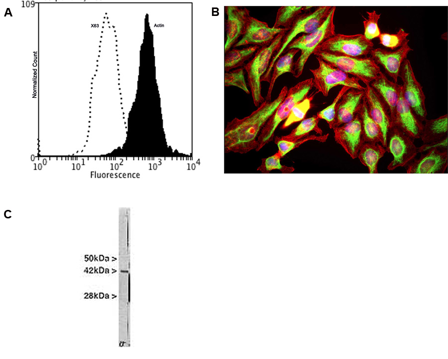
A: Specific staining of Actin expressed in human neuroblastoma SH-SY5Y cell line by Flow Cytometry using cat # M-1646-100. Fixing and Permeabilization of cells: Absolute methanol (10 minutes in ice) and 0.1% Tween-20 in PBS, Blocking: 1% BSA, Primary antibody: M-1646-100, 2 μg per ~10^6 cells, 30 minutes at room temperature, Secondary antibody: Goat anti-mouse PE (1:100), 20 minutes in dark at room temperature. Negative control: Non-specific Control IgG, clone X63 (cat # M-1249-200, black dashed). Data and results were generated using Orflo MoxiflowTM instrument and protocols. B: HeLa cells stained with M-1646-100 (red) and also with our chicken polyclonal antibody to Vimentin (C-1409-50). The actin antibody stains the submembranous actin rich cytoskeleton and also stress fibers, bundles of actin associated with adhesion sites. The vimentin antibody stains a quite different cytoskeletal network, the intermediate filaments. The blue stain reveals DNA in the nuclei of these cells. C: Crude extract of HeLa cells. The antibody recognizes the ~42 kDa protein.
Left: Actin expression in HeLa cells visualized with mouse antibody to actin (green) by Immunocytochemistry. Vimentin was co-stained with chicken antibody to vimentin (C-1409-50, red). Blue: DAPI nuclear stain.The actin antibody labels the submembranous actin-rich cytoskeleton, stress fibers and bundles of actin associated with cell adhesion sites. The vimentin antibody stains a different cytoskeletal network, the intermediate or 10 nm filaments. Right: Western blot analysis of tissue and cell lysates probed with mouse anti-actin antibody (red, lanes 2-7). [1] protein standard, [2] rat brain, [3] mouse brain, [4] NIH-3T3, [5] HEK293, [6] HeLa, [7] SH-SY5Y cells. The same blot was simultaneously probed chicken antibody against UCHL1 (C-1406-50, green), a marker of neuronal lineage cells.
General ReferencesVandekerckhove, J. and Weber, K. At least six different actins are expressed in a higher mammal: an analysis based on the amino acid. sequence of the amino-terminal tryptic peptide. J. Mol. Biol. 126:783-802 (1978).
 1800 605-5127
 +61 (0)8 8352 7711