 1800 605-5127
 +61 (0)8 8352 7711
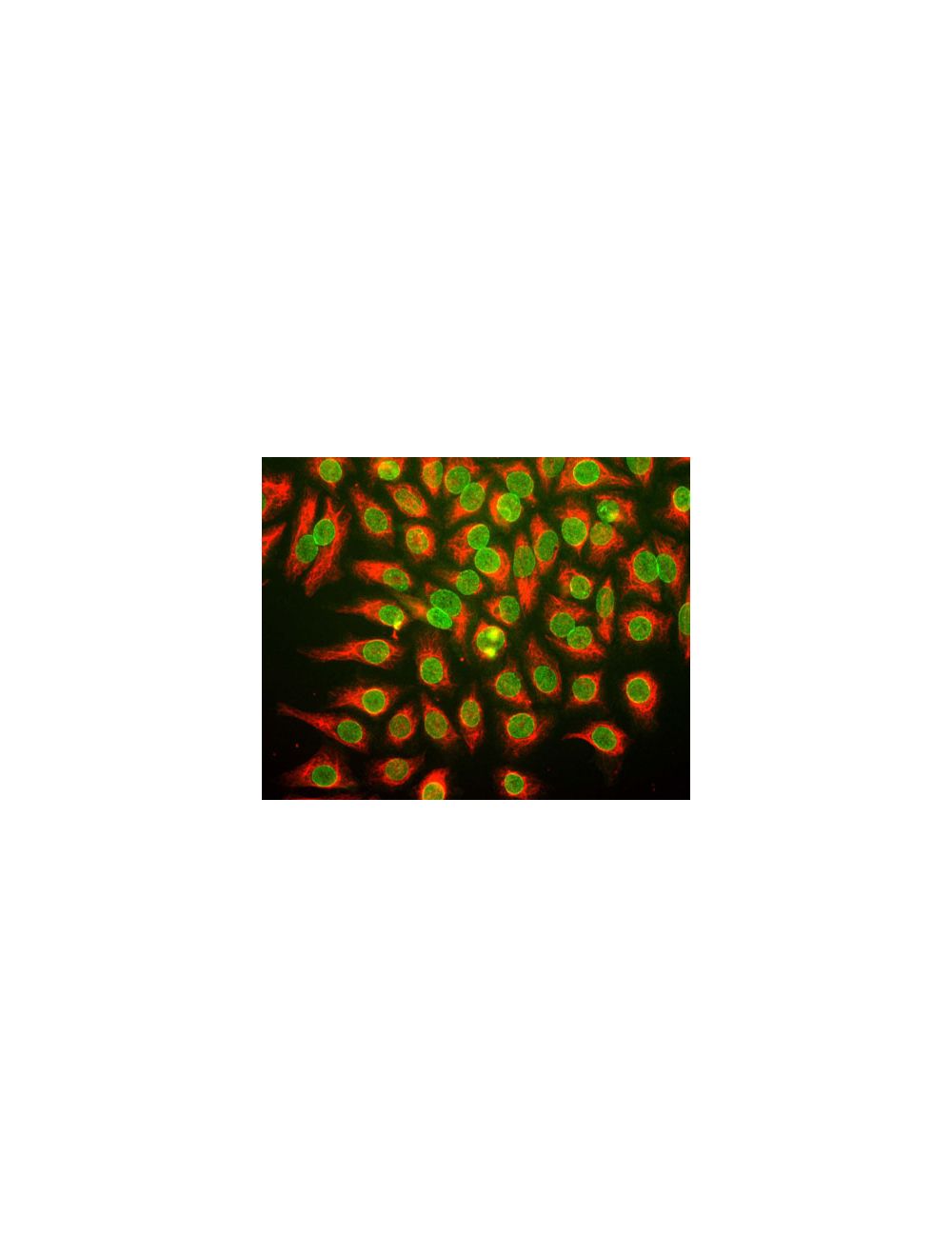

Nuclear pore complex protein Nup107, Mouse Monoclonal Antibody
- Product Name Nuclear pore complex protein Nup107, Mouse Monoclonal Antibody
-
Product Description
Mouse anti-Nuclear pore complex protein Nup107 Monoclonal Antibody (Unconjugated), suitable for ICC.
- Alternative Names Nuclear pore complex protein Nup107; 107 kDa nucleoporin; Nucleoporin Nup107; NUP107;
- Application(s) ICC
- Antibody Host Mouse
- Antibody Type Monoclonal
- Specificity The specificity of this antibody has been confirmed by IC. Hu, Rat, Ms, Yeast
- Species Reactivity Human, Mouse, Rat, Yeast
- Immunogen Description Yeast nuclear preparation
- Conjugate Unconjugated
- Purity Description Concentrated cell culture supernatant.
- Regulatory Status For research use only.
Product Info
-
Product Description
Mouse anti-Nuclear pore complex protein Nup107 Monoclonal Antibody (Unconjugated), suitable for ICC.
- Application(s) ICC
- Application Details Immunocytochemistry (ICC). A dilution of 1:50-1:500 is recommended for IC. This antibody does not work well for Western Blotting. Biosensis recommends optimal dilutions/concentrations should be determined by the end user.
- Target Nuclear pore complex protein Nup107
- Specificity The specificity of this antibody has been confirmed by IC. Hu, Rat, Ms, Yeast
- Target Host Species Yeast
- Species Reactivity Human, Mouse, Rat, Yeast
- Antibody Host Mouse
- Antibody Type Monoclonal
- Antibody Isotype IgG1
- Clone Name 39C7
- Conjugate Unconjugated
- Immunogen Description Yeast nuclear preparation
- Purity Description Concentrated cell culture supernatant.
- Format Lyophilized hybridoma cell culture media with sodium azide
- Reconstitution Instructions Spin the vial briefly before opening it. Reconstitute in 250 uL sterile-filtered, room temperature, ultrapure water. Let the vial rehydrate for 5 minutes. Mix gently with a pipette tip to help the redissolution of the material. Centrifuge to remove any insoluble material. The final solution will contain no preservatives. A sterile technique is recommended.
- Storage Instructions After reconstitution of lyophilized antibody, aliquot and store at -20°C for a higher stability. Avoid freeze-thaw cycles.
- Batch Number Please see item label.
- Expiration Date 12 months after date of receipt (unopened vial).
- Alternative Names Nuclear pore complex protein Nup107; 107 kDa nucleoporin; Nucleoporin Nup107; NUP107;
- Uniprot Number P57740
- Uniprot Number/Name 57740 (NU107_HUMAN)
- Scientific Background The Nuclear Core Complex (NPC) acts as a gateway for macromolecular traffic between the cytoplasm and the nucleus.
- Shipping Temperature 25°C (ambient)
- UNSPSC CODE 41116161
- Regulatory Status For research use only.

