Product DescriptionMouse anti-Splicing factor 3B subunit 4 (SF3B4) Monoclonal Antibody (Unconjugated), suitable for WB, ICC, FC.
Application(s)FC, ICC, WB
Application DetailsWB, ICC, Flow Cytometry. Recommended dilution of 1:500-1:2,000 for ICC. In WB using chemiluminescence it can be used at dilutions of 1:1,000 or lower. The protein runs on SDS-PAGE gels at an apparent molecular weight of 49 kDa. Use 2 ug/10^6 cells for Flow Cytometry. Biosensis recommends optimal dilutions/concentrations should be determined by the end user.
TargetSplicing factor 3B subunit 4 (SF3B4)
SpecificityHuman SF3B4 ; Bovine; Porcine; Mouse; Rat; expected to react with other species due to sequence homology
Target Host SpeciesHuman
Species ReactivityBovine, Human, Mouse, Other Mammals (Predicted), Pig, Rat
Antibody HostMouse
Antibody TypeMonoclonal
Antibody IsotypeIgG2b
Clone Name3A1
ConjugateUnconjugated
Immunogen DescriptionFull length recombinant human SF3B4 which was expressed in and purified from E. coli.
Purity DescriptionProtein G purified
FormatLyophilized from PBS buffer pH 7.2-7.6 with 0.1% trehalose, and sodium azide
Reconstitution InstructionsSpin vial briefly before opening. Reconstitute with 100 µL sterile-filtered, ultrapure water to achieve a 1 mg/mL concentration. Centrifuge to remove any insoluble material.
Storage InstructionsAliquot and store at -20°C for a higher stability and at 2-8°C with an appropriate antibacterial agent. Avoid freeze-thaw cycles.
Batch NumberPlease see item label.
Expiration Date12 months after date of receipt (unopened vial).
Alternative NamesSAP49; splicing factor 3b subunit 4; 49 kDa SAP49; spliceosome-associated protein 49; U2 snRNP; Hsh49; MGC108282; SF3B4; SF3b50;
Scientific BackgroundSF3B4 is one of 8 subunits of splicing factor SF3B. SF3B4 is ubiquitously expressed in the nuclei of eukaryotic cells, although it migrates into the cytoplasm of dividing cells.
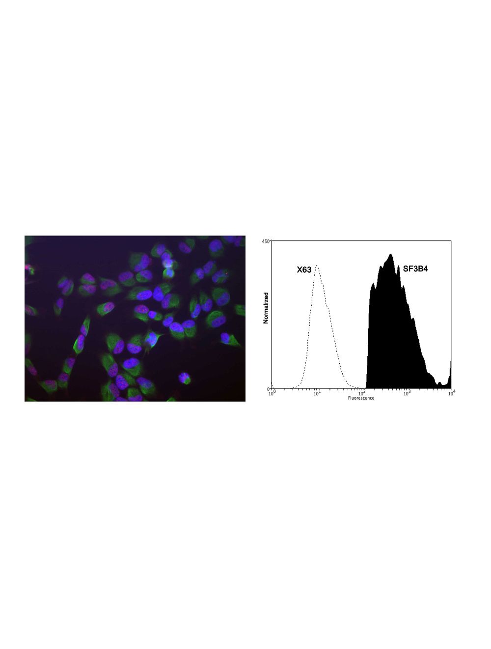
Left: Human HeLa cells stained with Mouse monoclonal antibody to splicing factor SF3B4 M-1576-100 (red), Chicken polyclonal antibody to Vimentin C-1409-50 (green) and DNA (blue, stained with DAPI). The monoclonal SF3B4 antibody reveals strong granular nuclear staining which is a little different from the DNA stain and presumably reflects splicosomal complexes. The polyclonal Vimentin antibody stains the cytoplasmic intermediate filament network of the HeLa cells. Right: Analysis of SF3B4 expression in rat pheochromocytoma PC-12 cell line by Flow Cytometry. Fixing and Permeabilization of cells: Absolute methanol (10 minutes in ice) and 0.1% Tween-20 in PBS, Blocking: 1% BSA, Primary antibody: Mouse Monoclonal antibody to SF3B4 (cat # M-1576-100, 2μg per ~10^6 cells) for 30 minutes at room temperature, Secondary antibody: Goat anti-mouse PE labeled secondary antibody (1:100 fold dilution) with incubation for 20 minutes in dark at room temperature. Non-specific Control IgG, clone X63 (cat # M-1249-100) was used as negative control under same conditions (black dashed). Flow cytometry data and results were generated using Orflo MoxiflowTM instrument and protocols.
Left: Analysis of SF3B4 expression in HeLa cells by Immunocytochemistry. Cells were stained with mouse antibody to splicing factor SF3B4 (red, 1:1,000), and co-stained with chicken antibody to vimentin (C-1409-50, green, 1:10,000). Blue: DAPI nuclear stain. The SF3B4 antibody reveals strong granular staining of the nuclei, while the vimentin antibody specifically labels cytoplasmic intermediate filaments. Right: Western blot analysis of cell lysates, cytosol- or nuclear-enriched fractions, using mouse antibody to splicing factor SF3B4 (green, 1:1,000). [1] protein standard, [2] NIH-3T3 cytosolic fraction, [3] NIH-3T3 nuclear fraction, [4] HeLa cytosolic and [5] HeLa nuclear fractions. A strong single band at 49 kDa represents the SF3B4 protein, which is expressed exclusively in the nuclei. The same blot was simultaneously probed with rabbit anti-GAPDH antibody (R-1701-100, red, 1:20,000, lanes 2-5). The 37 kDa band corresponds to the GAPDH protein, detected mainly in the cytosolic fractions of these cells.
 1800 605-5127
 +61 (0)8 8352 7711