Product NameNeurofilament medium polypeptide (NF-M), Mouse Monoclonal Antibody (3H11)
Product DescriptiongoogleMouse anti-Neurofilament medium polypeptide (NF-M), Monoclonal Antibody (Unconjugated), suitable for WB, Immunostaining and FC.
Alternative NamesNF-M; NFM; Neurofilament medium polypeptide; 160 kDa neurofilament protein; Neurofilament 3; Neurofilament triplet M protein; Nefm; Nef3; Nfm
Application(s)FC, IF, ICC, IHC, WB
Antibody HostMouse
Antibody TypeMonoclonal
SpecificitySpecies cross-reactivity includes human, rat, mouse, cow, pig, horse and chicken.
Species ReactivityBovine, Chicken, Horse, Human, Mouse, Pig, Rat
Immunogen DescriptionThis antibody has been made against a recombinant fusion protein containing the extreme C-terminus of rat NF-M (amino acids 677-845) expressed in and purified from E. coli. The epitope is localized to within the last 56 amino acids at the extreme C-terminus of rat NF-M, the so-called KE segment which is highly conserved between NF-M molecules from different species.
Application DetailsWestern blot (WB), Immunocytochemistry (ICC) / Immunofluorescence (IF), Immunohistochemistry (IHC) and Flow Cytometry (FC). A dilution of 1:5,000 is recommended for WB. A dilution of 1:2,000 is recommended for ICC/IFF and IHC. A dilution of 2 ug per 10^6 cells is recommended for FC. Biosensis recommends optimal dilutions/concentrations should be determined by the end user.
TargetNeurofilament medium polypeptide (NF-M)
SpecificitySpecies cross-reactivity includes human, rat, mouse, cow, pig, horse and chicken.
Target Host SpeciesRat
Species ReactivityBovine, Chicken, Horse, Human, Mouse, Pig, Rat
Antibody HostMouse
Antibody TypeMonoclonal
Antibody IsotypeIgG1
Clone Name3H11
ConjugateUnconjugated
Immunogen DescriptionThis antibody has been made against a recombinant fusion protein containing the extreme C-terminus of rat NF-M (amino acids 677-845) expressed in and purified from E. coli. The epitope is localized to within the last 56 amino acids at the extreme C-terminus of rat NF-M, the so-called KE segment which is highly conserved between NF-M molecules from different species.
Purity DescriptionProtein G purified
FormatLyophilized from PBS buffer pH 7.2-7.6 with 0.1% trehalose, and sodium azide
Reconstitution InstructionsSpin vial briefly before opening. Reconstitute with 100 µL sterile-filtered, ultrapure water to achieve a 1 mg/mL concentration. Centrifuge to remove any insoluble material.
Storage InstructionsStore lyophilized antibody at 2-8°C After reconstitution of lyophilized antibody, aliquot and store at -20°C for a higher stability. Avoid freeze-thaw cycles. Store at 4°C for up to one month for short term storage and frequent use.
Batch NumberPlease see item label.
Expiration Date12 months after date of receipt (unopened vial).
Alternative NamesNF-M; NFM; Neurofilament medium polypeptide; 160 kDa neurofilament protein; Neurofilament 3; Neurofilament triplet M protein; Nefm; Nef3; Nfm
Scientific BackgroundNeurofilaments are the 10nm or intermediate filament proteins found specifically in neurons, and are composed predominantly of three major proteins called NF-L, NF-M and NF-H, though other filament proteins may be included also. The major function of neurofilaments is likely to control the diameter of large axons. NF-L is the neurofilament light or low molecular weight polypeptide and runs on SDS-PAGE gels at 68-70kDa with some variability across species. Antibodies to NF-L are useful for identifying neuronal cells and their processes in cell culture and sectioned material. NF-L antibody can also be useful for the visualization of neurofilament rich accumulations seen in many neurological diseases, such as Lou Gehrig’s disease (ALS), giant axon neuropathy, Charcot-Marie Tooth disease and others. (Ref: uniprot.org)
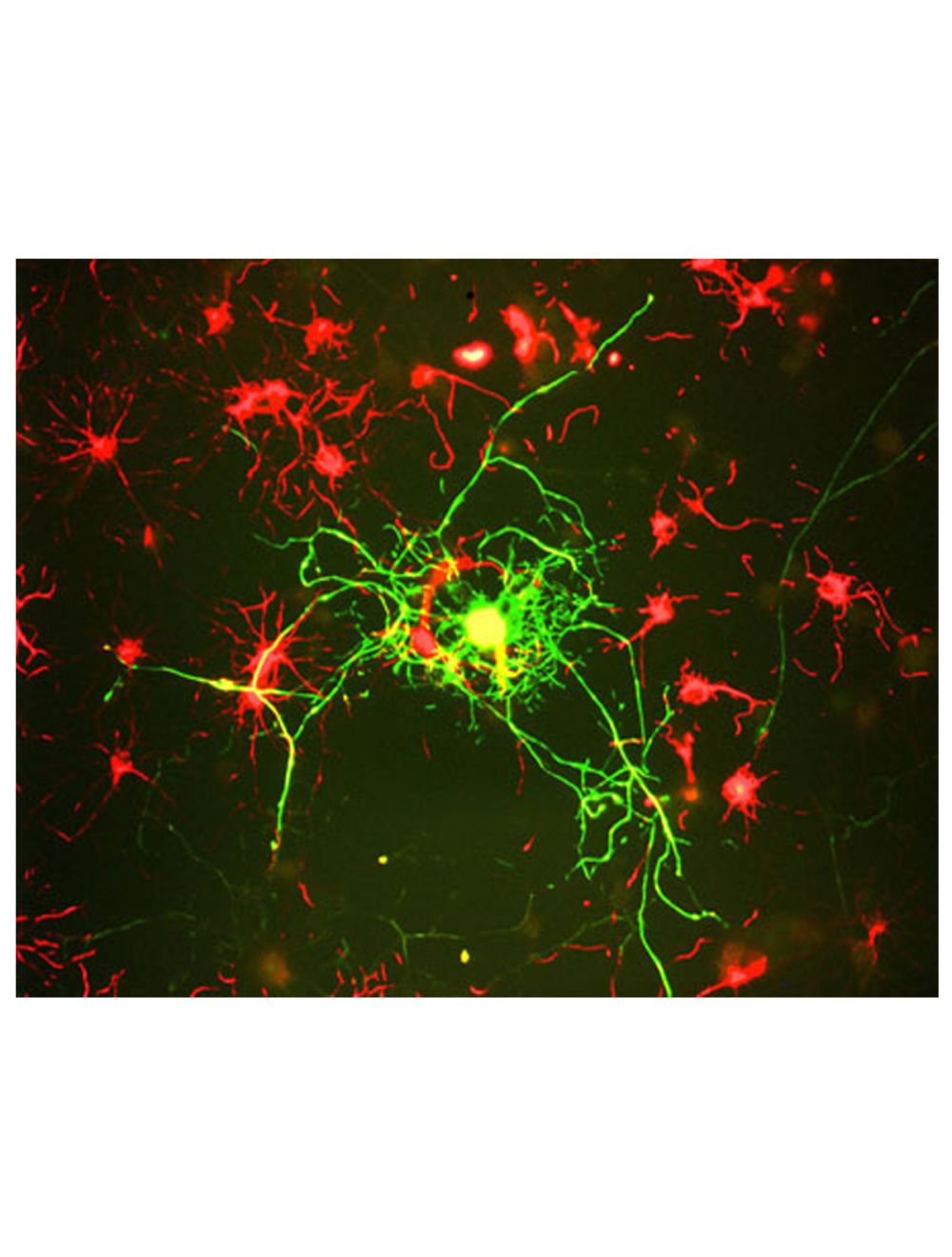
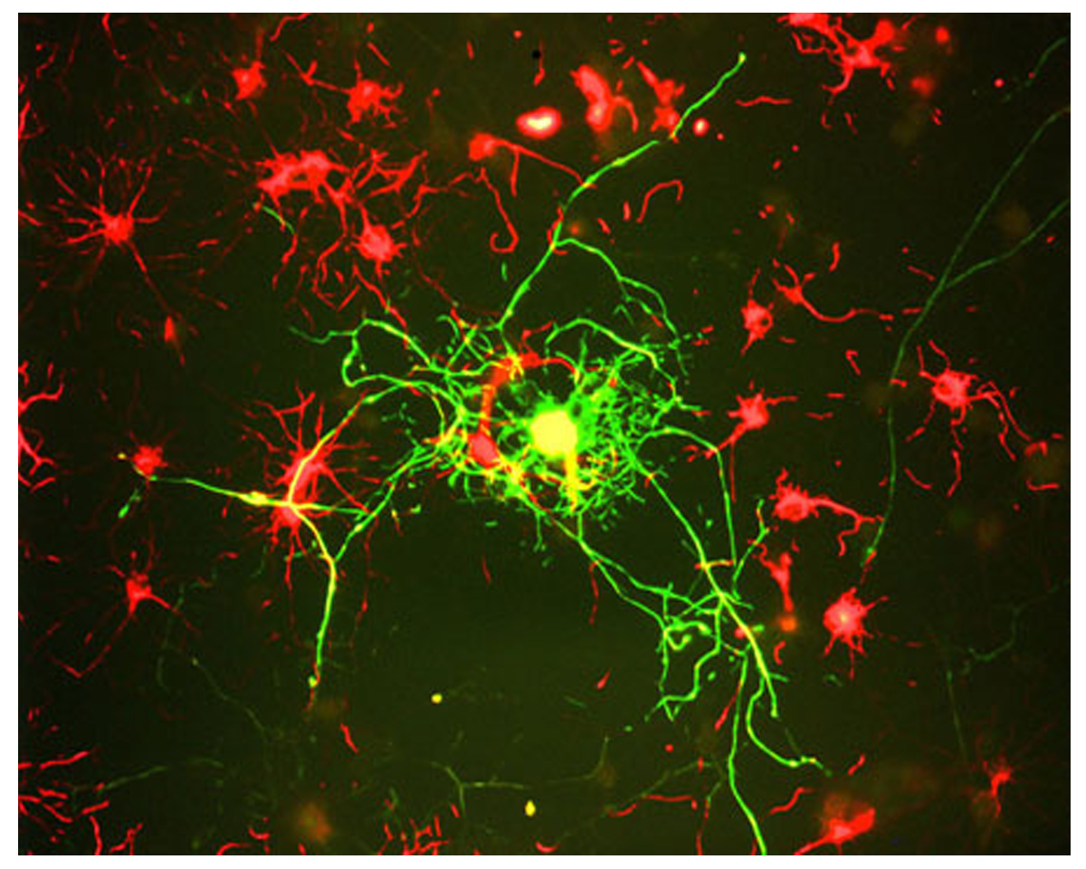
Image of a neuron and surrounding cells in cerebral culture, derived from adult rats, by Immunofluorescence and Immunocytochemistry. The cells were stained with M-1394-100, Neurofilament medium polypeptide (NF-M), Clone 3H11, Mouse mAb, (green), and co-stained with product R-1379-50, Internexin alpha, Rabbit pAb, (red). Mature neurons can be identified by their morphology and because they stain strongly with antibodies to NF-L, NF-M and NF-H (green). The surrounding mitotic neuronal progenitor cells (red) express many other neuronal markers.
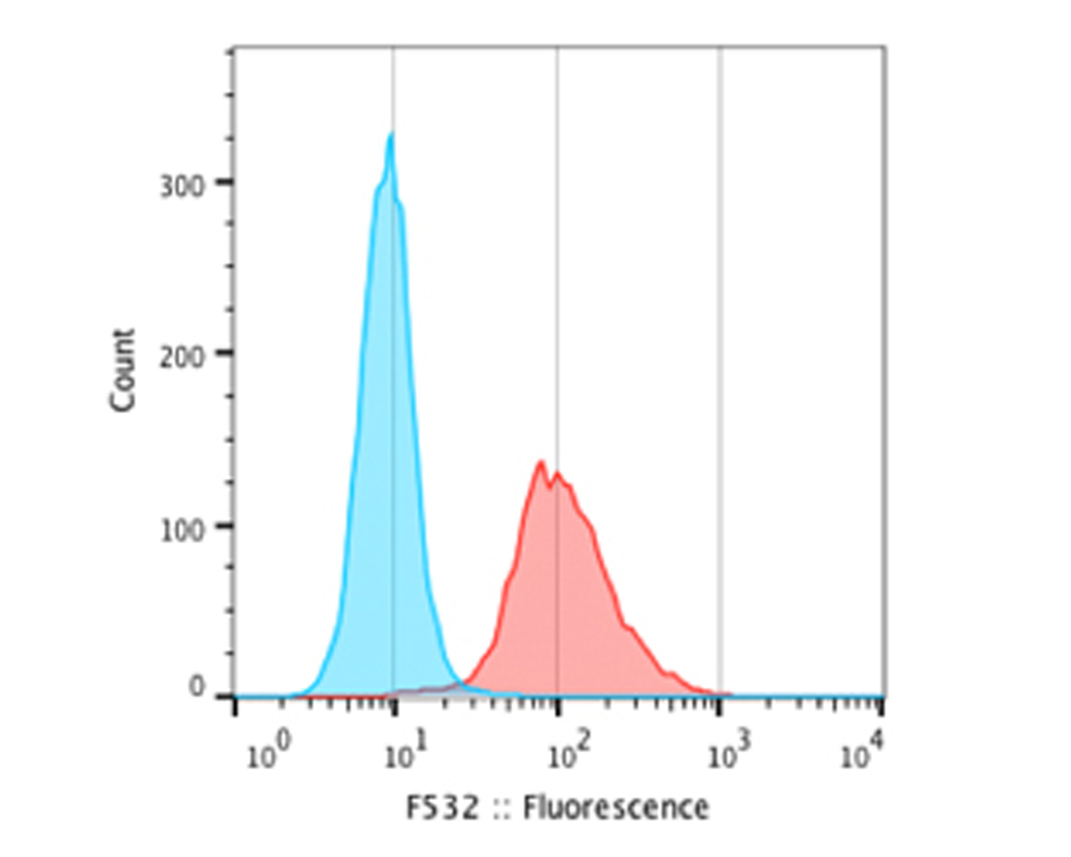
Analysis by flow cytometry of Neurofilament medium polypeptide (NF-M), (endogenous) expression in mouse neural progenitor cells differentiated from mES and fixed overnight in 70% ethanol (red curve). PE-labelled goat anti-mouse IgG was used as secondary antibody (Blue curve) and negative control processed with secondary antibody only. Flow cytometry data and results were generated using Orflo MoxiflowTM instrument and protocols.
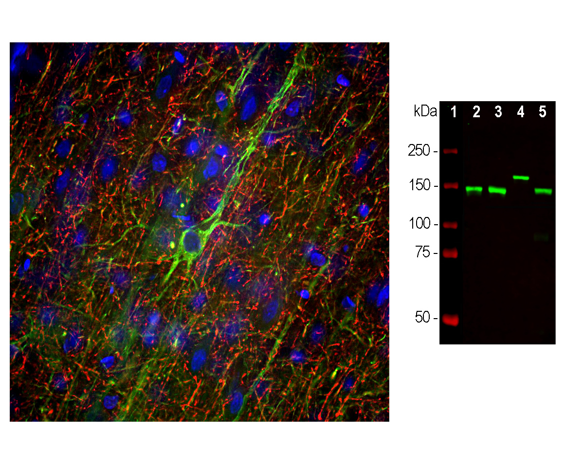
Left: Analysis by Immunofluorescence and Immunohistochemistry of Neurofilament medium polypeptide (NF-M) expression in rat frontal cortex section. Section was stained with M-1394-100, Neurofilament medium polypeptide (NF-M), Clone 3H11, Mouse mAb, (green, 1:5000), and co-stained with product C-1386-50, Neurofilament heavy polypeptide (NF-H), Chicken pAb (red, 1:5000). IHC Method: Following transcardial perfusion of rat with 4% paraformaldehyde, brain was post-fixed for 24 hours, cut into 45 um sections, and free-floating sections were stained. The NF-M mouse antibody labels neuronal cell bodies and dendrites of pyramidal neurons, as well as dendrites and axons of other neuronal cells, while the chicken NF-H antibody stains the network of neuronal axons only. Right: Analysis by western blot of NF-M expression in neuronal tissue lysates using M-1394-100 (green, 1:10,000) with a band at approx 145kDa (rodent NF-M) and 160kDa (bovine NF-M). Lane 1: protein standard; Lane 2: rat spinal cord; Lane 3: mouse spinal cord; Lane 4: cow spinal cord; Lane 5: rat sciatic nerve.
Specific ReferencesFelitsyn N. et al (2008) The heme precursor delta-aminolevulinate blocks peripheral myelin formation. J Neurochem. 2008 Sep;106(5):2068-79.
 1800 605-5127
 +61 (0)8 8352 7711