 1800 605-5127
 +61 (0)8 8352 7711
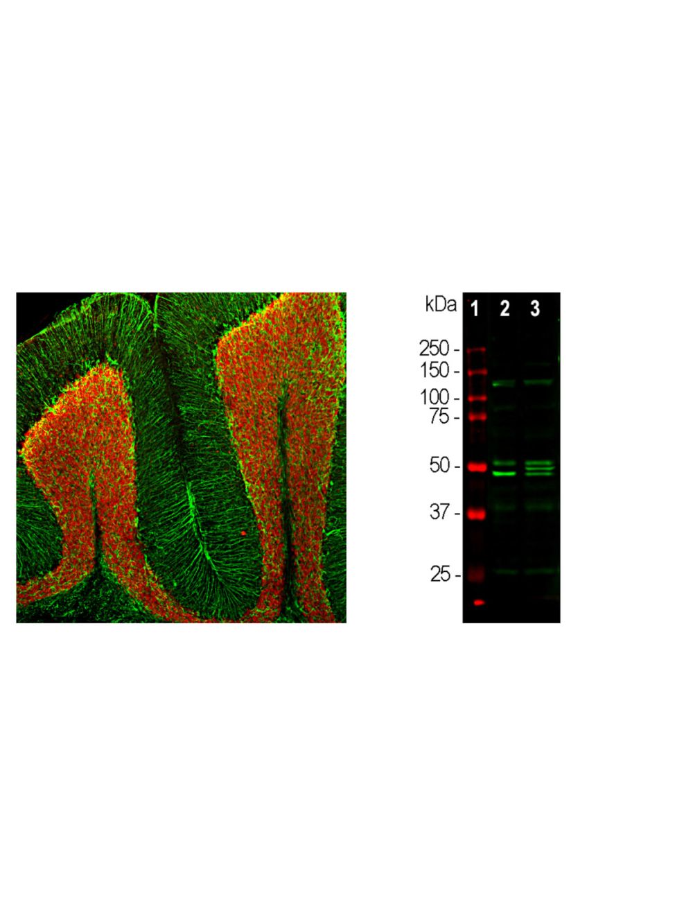

Neuronal nuclei antigen/Fox3 (NeuN/Fox3), Rabbit Polyclonal Antibody
- Product Name Neuronal nuclei antigen/Fox3 (NeuN/Fox3), Rabbit Polyclonal Antibody
- Product Description Rabbit anti-Neuronal nuclei antigen/Fox3 (NeuN/Fox3) Polyclonal Antibody (Unconjugated), suitable for WB, IHC-Frozen, ICC.
-
Replacement Product
Replaced by R-3771-100.
- Alternative Names Feminizing locus on X; Fox-1; Fox3; NeuN;
- Application(s) ICC, IHC-Frozen, WB
- Antibody Host Rabbit
- Antibody Type Polyclonal
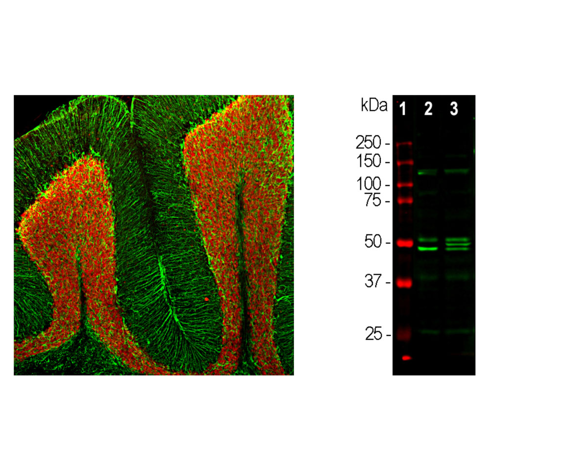
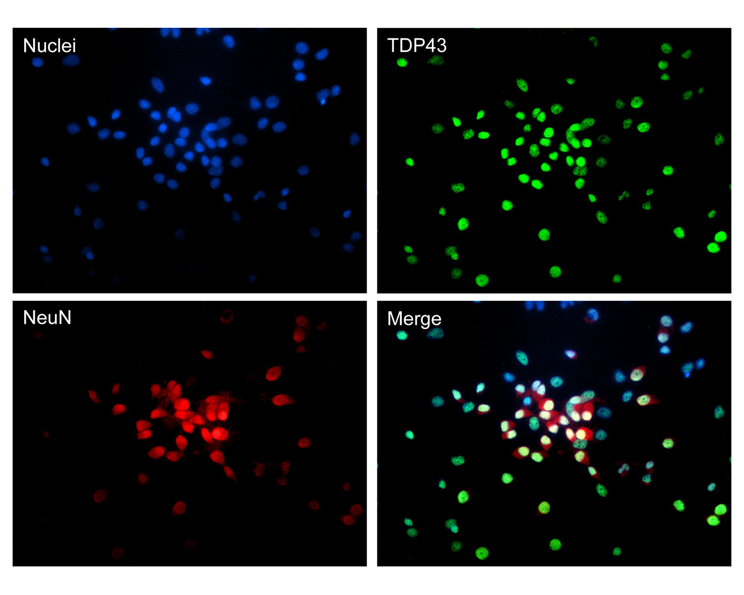
- Specificity The specificity of this antibody has been confirmed by WB and IC. Two alternate transcripts can be seen at 46 kDa and 48 kDa in WB. This antibody has been used successfully for immunohistochemistry on paraformaldehyde fixed rat brain cortex (adult) tissue. It does not bind to the nuclei of perikarya of non-neuronal cells making it a good marker to identify neurons. Rat. Predicted to react with other mammals due to sequence homology.
- Species Reactivity Human, Mouse, Rat
- Immunogen Description Antibody was raised against the N-terminal 100 amino acids of human Fox3 as expressed in and purified from E. coli.
- Conjugate Unconjugated
- Purity Description Whole serum
- Regulatory Status For research use only.
Product Info
- Product Description Rabbit anti-Neuronal nuclei antigen/Fox3 (NeuN/Fox3) Polyclonal Antibody (Unconjugated), suitable for WB, IHC-Frozen, ICC.
-
Replacement Product
Replaced by R-3771-100.
- Application(s) ICC, IHC-Frozen, WB
- Application Details Western Blotting (WB), Immunocytochemistry (ICC) and Immunohistochemistry (IHC). A dilution of 1:1,000 - 1:2,000 is recommended for WB. A dilution of 1:500 - 1:1,000 is recommended for ICC and IHC. Biosensis recommends optimal dilutions/concentrations should be determined by the end user.
- Target Neuronal nuclei antigen/Fox3 (NeuN/Fox3)
- Specificity The specificity of this antibody has been confirmed by WB and IC. Two alternate transcripts can be seen at 46 kDa and 48 kDa in WB. This antibody has been used successfully for immunohistochemistry on paraformaldehyde fixed rat brain cortex (adult) tissue. It does not bind to the nuclei of perikarya of non-neuronal cells making it a good marker to identify neurons. Rat. Predicted to react with other mammals due to sequence homology.
- Target Host Species Human
- Species Reactivity Human, Mouse, Rat
- Antibody Host Rabbit
- Antibody Type Polyclonal
- Antibody Isotype Mixed
- Conjugate Unconjugated
- Immunogen Description Antibody was raised against the N-terminal 100 amino acids of human Fox3 as expressed in and purified from E. coli.
- Purity Description Whole serum
- Format Lyophilized with sodium azide.
- Reconstitution Instructions Spin vial briefly before opening. Reconstitute with 100 µL sterile-filtered, ultrapure water. Centrifuge to remove any insoluble material.
- Storage Instructions After reconstitution of lyophilized antibody, aliquot and store at -20°C for a higher stability. Avoid freeze-thaw cycles.
- Batch Number Please see item label.
- Expiration Date 12 months after date of receipt (unopened vial).
- Alternative Names Feminizing locus on X; Fox-1; Fox3; NeuN;
- Uniprot Number A6NFN3
- Uniprot Number/Name A6NFN3 (RFOX3_HUMAN)
- Scientific Background Fox3 is one of a family of mammalian homologues of Fox-1. The Fox proteins are about 46 kDa in size, and each includes a central highly conserved RRM type RNA recognition motif. Much interest has focused on Fox3 as a result of the recent finding that this protein corresponds to NeuN, a neuronal nuclear antigen. NeuN/Fox-3 has a function in RNA splicing and is expressed heavily and specifically in neuronal nuclei and cytoplasm. Our antibody was raised against the N-terminal 100 amino acids of human Fox3 as expressed in and purified from E. coli. We did not use full length Fox3 as immunogen since the three mammalian Fox homologues, namely Fox1, Fox2 and Fox3, include virtually identical RRM motifs. The N-terminal region of the three molecules are much more variable in the three molecules so antibodies specific for each of the three molecules can therefore be generated.
- Shipping Temperature 25°C (ambient)
- UNSPSC CODE 41116161
- Regulatory Status For research use only.
Specifications


-
Specific References
Chun E (2021) Structural and Functional Consequences of Targeted Hippocampal Gaba Neuron Ablation by Stable Substance P-Saporin in Rats. PhD Thesis. Species: Rat. Application IHC/IF.
Roberts BM et al (2020) GABA uptake transporters support dopamine release in dorsal striatum with maladaptive downregulation in a parkinsonism model. Nat Commun. 11(1):4958 Species: Mouse. Application IHC/IF.
Kumamaru H et al (2019) Regenerating Corticospinal Axons Innervate Phenotypically Appropriate Neurons within Neural Stem Cell Grafts. Cell Rep. 26(9):2329-39 Species: Rat, monkey. Application IHC/IF.
Castle MJ et al (2018) Physical positioning markedly enhances brain transduction after intrathecal AAV9 infusion. Sci Adv. 4(11):eaau9859 Species: Rat. Application IHC/IF.
Hao XZ et al (2016) Combining systemic and stereotactic MEMRI to detect the correlation between gliosis and neuronal connective pathway at the chronic stage after stroke. J Neuroinflammation. 13(1):156 Species: Rat. Application IH.
Zhou XF et al (2015) Transcription factors COUP-TFI and COUP-TFII are required for the production of granule cells in the mouse olfactory bulb.Development. 142(9):1593-605 Species: Mouse. Application IH.
Saha M, et al (2013) Spinal Mitogen-Activated Protein Kinase Phosphatase-3 (MKP-3) is Necessary for the Normal Resolution of Mechanical Allodynia in a Mouse Model of Acute Postoperative Pain. J. Neurosci. 33(43):17182-7
Liu X et al (2013) Suppressive Effect of Phenol Red on the Epileptiform Burst Activity via Activation of Estrogen Receptors in Primary Hippocampal Culture. PLoS ONE 8(4):e60189
Landry R.P. et al (2012) Spinal Cannabinoid Receptor Type 2 Agonist Reduces Mechanical Allodynia and Induces Mitogen-Activated Protein Kinase Phosphatases in a Rat Model of Neuropathic Pain. J Pain. 2012 Aug 14
Ndong C. et al (2012) Mitogen activated protein kinase phosphatase-1 prevents the development of tactile sensitivity in a rodent model of neuropathic pain. Mol Pain. 2012 Apr 27;8(1):34.