SpecificityThe specificity of this antibody has been confirmed by WB. This antibody detects ~24 kDa UCHL1 enzyme. Suitable control tissue is rat spinal cord or peripheral nerve homogenate. Hu, Rat, Ms, Bov, Por. Predicted to react with other mammalian tissues due to sequence homology.
Species ReactivityBovine, Human, Mouse, Other Mammals (Predicted), Pig, Rat
Immunogen DescriptionRecombinant full length human Ubiquitin C Terminal Hydrolase 1 (UCHL1) purified from E. coli.
Application DetailsWestern Blotting (WB) and Immunocytochemistry (ICC). A dilution of 1:5,000 - 1:10,000 is recommended for WB. A dilution of 1:500-1,000 is recommended for IC. Biosensis recommends optimal dilutions/concentrations should be determined by the end user.
SpecificityThe specificity of this antibody has been confirmed by WB. This antibody detects ~24 kDa UCHL1 enzyme. Suitable control tissue is rat spinal cord or peripheral nerve homogenate. Hu, Rat, Ms, Bov, Por. Predicted to react with other mammalian tissues due to sequence homology.
Target Host SpeciesHuman
Species ReactivityBovine, Human, Mouse, Other Mammals (Predicted), Pig, Rat
Antibody HostChicken
Antibody TypePolyclonal
Antibody IsotypeIgY
ConjugateUnconjugated
Immunogen DescriptionRecombinant full length human Ubiquitin C Terminal Hydrolase 1 (UCHL1) purified from E. coli.
Purity DescriptionIgY
FormatLyophilized IgY preparation, with sodium azide.
Reconstitution InstructionsSpin vial briefly before opening. Reconstitute with 50 µL sterile-filtered, ultrapure water. Centrifuge to remove any insoluble material.
Storage InstructionsAfter reconstitution of lyophilized antibody, aliquot and store at -20°C for a higher stability. Avoid freeze-thaw cycles.
Batch NumberPlease see item label.
Expiration Date12 months after date of receipt (unopened vial).
Alternative NamesUbiquitin carboxyl-terminal hydrolase isozyme L1; UCH-L1; Neuron cytoplasmic protein 9.5; PGP 9.5; PGP9.5; Ubiquitin thioesterase L1; UCHL1; Ubiquitin C Terminal Hydrolase 1;
Scientific BackgroundThis enzyme is a thiol protease that recognizes and hydrolyzes a peptide bond at the C-terminal glycine of ubiquitin. The enzyme also binds to free monoubiquitin and may prevent its degradation in lysosomes (ref: SWISSPROT).
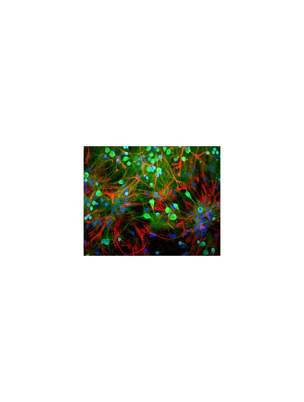
Image shows rat mixed neuron/glial cultures stained with Chicken polyclonal antibody to Ubiquitin C Terminal Hydrolase 1 C-1406-50 (green) and Rabbit polyclonal antibody to Glial Fibrillary Acidic Protein R-1374-50 (red). Blue is a DNA stain. Note that the Ubiquitin antibody stains neurons strongly and specifically and that the staining is concentrated in the cell bodies, though some does extend into the dendrites also.
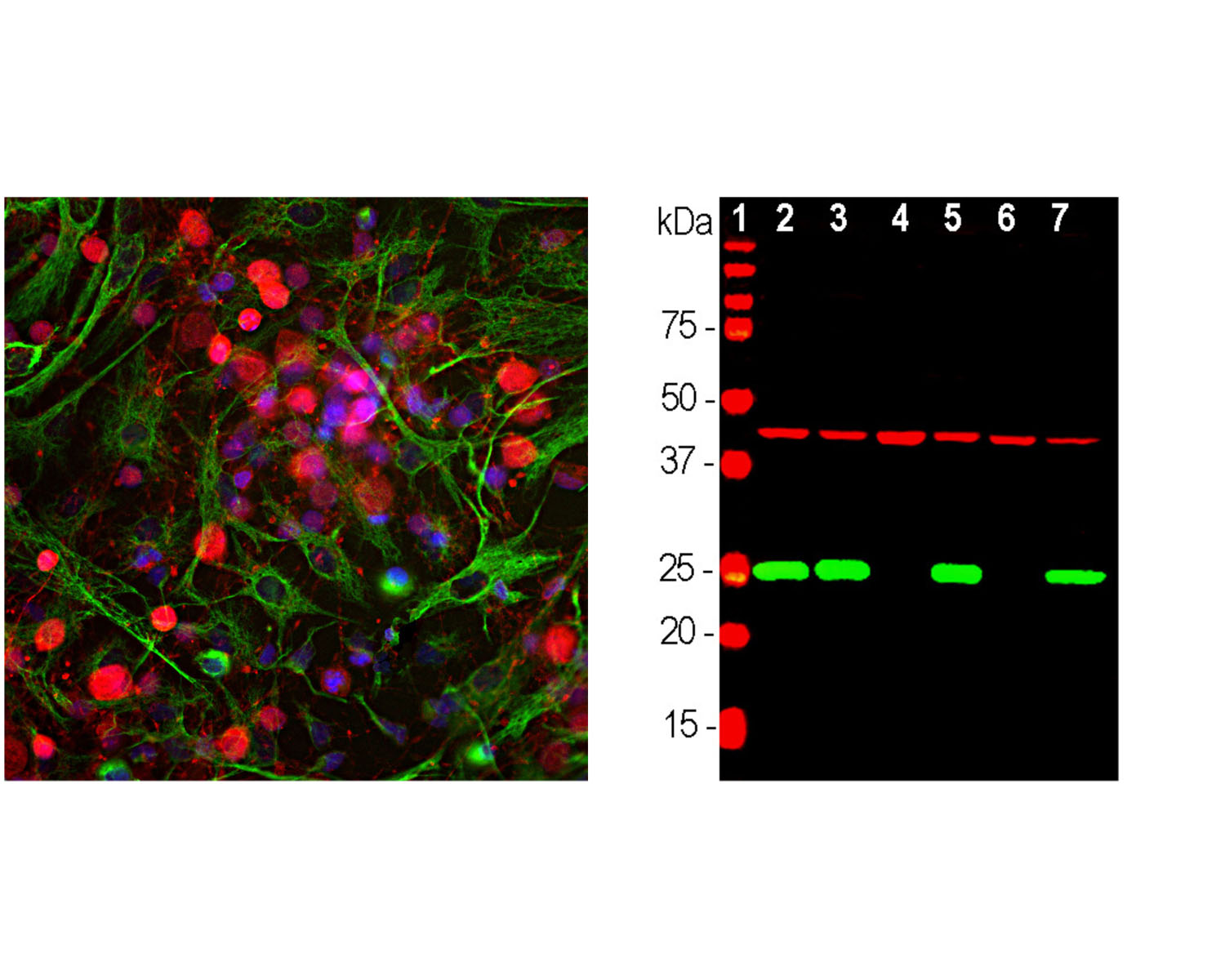
Left: Analysis of UCHL1 expression in cortical neuron-glial cell culture from E20 rat by Immunocytochemistry with chicken antibody to UCHL1 (1:500, red). Cells were co-stained with a mouse antibody to vimentin (green). Blue: DAPI nuclear stain. The UCHL1 antibody produces strong staining of the cell body and dendrites in neurons. The vimentin antibody stains intermediate filaments in fibroblastic and developing glial cells. Right: Western blot analysis of tissue and cell lysates using chicken antibody to UCHL1 (1:2,000, green), and a mouse anti-actin antibody (red, lanes 2-7). [1] protein standard, [2] rat brain, [3] mouse brain, [4] NIH-3T3, [5] HEK293, [6] HeLa and [7] SH-SY5Y cells. The single band at 24 kDa mark corresponds to UCHL1 protein which is detectable in CNS extracts and lysates of cells with neuronal properties, but not in lysates of HeLa, NIH-3T3 and other non-neuronal cells. Actin is detected with apparent molecular weight of 42 kDa and provides an excellent loading control.
 1800 605-5127
 +61 (0)8 8352 7711