Application DetailsWestern blot: 0.1-0.5 μg/mL. Immunohistochemistry (paraffin-embedded sections): 0.5-1 μg/mL, heat-mediated antigen-retrieval required. Immunohistochemistry (frozen sections): 0.5-1 μg/mL. Immunocytochemistry: 2 μg/mL. Flow Cytometry: 1-3 μg/1x10^6 cells. Biosensis recommends optimal dilutions/concentrations should be determined by the end user.
Product ValidationValidated by IHC, WB, ICC and FC.
TargetCofilin-1 (CFL1)
SpecificityNo cross reactivity with other proteins.
Target Host SpeciesHuman
Species ReactivityHuman, Mouse, Primate, Rat
Antibody HostRabbit
Antibody TypePolyclonal
Antibody IsotypeIgG
ConjugateUnconjugated
Immunogen DescriptionE.coli-derived recombinant human Cofilin-1 protein (amino acid position: A2-L166).
Purity DescriptionPurified rabbit IgG.
Physical StateSolid
FormatLyophilized from a solution containing 5 mg BSA, 0.9 mg NaCl, 0.2 mg Na2HPO4, 0.05 mg NaN3.
Reconstitution InstructionsSpin vial briefly before opening. Reconstitute in 200 μL sterile-filtered, ultrapure water to achieve a concentration of 0.5 mg/mL. Centrifuge to remove any insoluble material.
Storage InstructionsStore lyophilized antibody at 2-8°C. After reconstitution divide into aliquots and store at -20°C for long-term storage. Store at 2-8°C short-term (up to 4 weeks). Avoid repetitive freeze/thaw cycles.
Batch NumberPlease see item label.
Expiration Date12 months after date of receipt (unopened vial).
Alternative Names18 kDa phosphoprotein; p18; Cofilin, non-muscle isoform
Scientific BackgroundBinds to F-actin and exhibits pH-sensitive F-actin depolymerizing activity (PubMed:11812157). In conjunction with the subcortical maternal complex (SCMC), plays an essential role for zygotes to progress beyond the first embryonic cell divisions via regulation of actin dynamics (PubMed:15580268). Required for the centralization of the mitotic spindle and symmetric division of zygotes (By similarity). Plays a role in the regulation of cell morphology and cytoskeletal organization in epithelial cells (PubMed:21834987). Required for the up-regulation of atypical chemokine receptor ACKR2 from endosomal compartment to cell membrane, increasing its efficiency in chemokine uptake and degradation (PubMed:23633677). Required for neural tube morphogenesis and neural crest cell migration (By similarity). Ref: uniprot.org
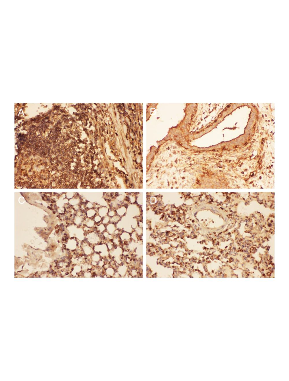
Analysis of Cofilin expression (brown) by Immunohistochemistry in paraffin-embedded sections of human intestinal cancer tissue (A), human mammary cancer tissue (B), mouse lung tissue (C), and rat lung tissue (D). Heat-mediated antigen retrieval was performed in citrate buffer (pH 6, epitope retrieval solution) for 20 minutes. The tissue sections were blocked with 10% goat serum, and then incubated with 1 μg/mL rabbit anti-cofilin antibody (overnight at 4°C). Biotinylated goat anti-rabbit IgG was used as secondary antibody and incubated for 30 minutes at 37°C. The tissue section was developed using Strepavidin-Biotin-Complex with DAB as the chromogen.
Analysis of Cofilin expression (brown) in frozen section of human placenta tissue by Immunohistochemistry. The tissue section was blocked with 10% goat serum, and then incubated with 1 μg/mL rabbit anti-cofilin Antibody (overnight at 4°C). Biotinylated goat anti-rabbit IgG was used as secondary antibody and incubated for 30 minutes at 37°C. The tissue section was developed using Strepavidin-Biotin-Complex with DAB as the chromogen.
Western blot analysis of Cofilin expression in cell lysates and tissue homogenates. Cofilin protein is detected at the expected molecular weight of ~18 kDa. SDS-PAGE: 50 µg protein per lane, reducing conditions. Western Blot: Transfer: nitrocellulose membrane; Blocking: 5% skim milk; Primary antibody: 0.5 µg/mL overnight; Secondary antibody: anti-rabbit-HRP conjugate; Detection: Chemiluminiscence.
Analysis of Cofilin expression (green) in U20S cells by Immunocytochemistry. Cells were blocked with 10% goat serum, prior to addition of 2 µg/mL dilution of rabbit anti-cofilin primary antibody (overnight incubation). A secondary green-fluorophore tagged antibody was used to visualize cofilin expression.
Analysis of Cofilin expression (blue) in U20S cells by Flow Cytometry. Cells were blocked with 10% goat serum and then incubated with rabbit anti-cofilin antibody (1 μg/1x106 cells) for 30 min. A green fluorophore-tagged secondary anti-rabbit antibody was used. Isotype control antibody (rabbit IgG, green line) and unlabelled sample (red line) were used as control.
 1800 605-5127
 +61 (0)8 8352 7711