1800 605-5127
 +61 (0)8 8352 7711
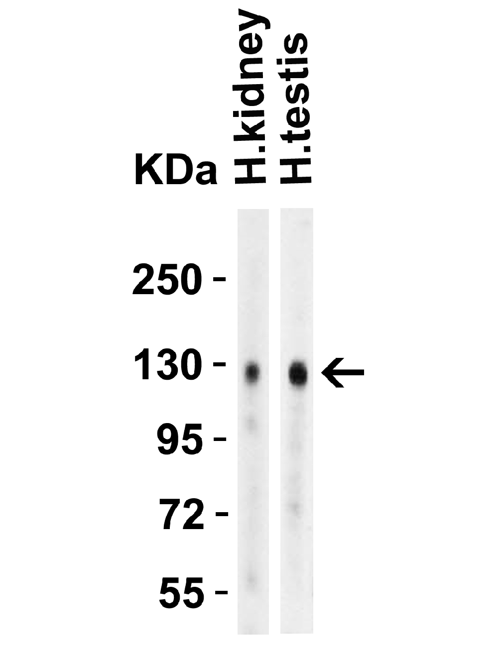

Angiotensin converting enzyme 2 (ACE2), Rabbit Polyclonal Antibody
- Product Name Angiotensin converting enzyme 2 (ACE2), Rabbit Polyclonal Antibody
- Product Description SARS-CoV Spike Antibody: A novel coronavirus has been identified as the causative agent of SARS (Severe Acute Respiratory Syndrome). Coronaviruses are a major cause of upper respiratory diseases in humans. The genomes of these viruses are positive-stranded RNA approximately 27-31kb in length. SARS infection can be mediated by the binding of the viral spike protein, a glycosylated 139 kDa protein and the major surface antigen of the virus, to the angiotensin-converting enzyme 2 (ACE2) on target cells. This binding can be blocked by a soluble form of ACE2.
- Alternative Names ACE2 Antibody: ACEH, Angiotensin-converting enzyme 2, ACE-related carboxypeptidase, ACEH, SARS-CoV receptor, SARS-CoV-2 receptor
- Application(s) ELISA, ICC, IHC-Paraffin-embedded, WB
- Antibody Host Rabbit
- Antibody Type Polyclonal
- Specificity Anti-ACE2 has no cross response to ACE1. Predicted species reactivity based on immunogen sequence: Bovine: (93%)
- Species Reactivity Human, Mouse, Rat
-
Immunogen Description
ACE2 antibody was raised against a synthetic peptide corresponding to amino acids near the center of human ACE2.
The immunogen is located within amino acids 150 - 200 of ACE2. - Conjugate Unconjugated
- Concentration 1 mg/mL
- Purity Description ACE2 Antibody is affinity chromatography purified via peptide column.
- Regulatory Status For research use only.
Product Info
- Product Description SARS-CoV Spike Antibody: A novel coronavirus has been identified as the causative agent of SARS (Severe Acute Respiratory Syndrome). Coronaviruses are a major cause of upper respiratory diseases in humans. The genomes of these viruses are positive-stranded RNA approximately 27-31kb in length. SARS infection can be mediated by the binding of the viral spike protein, a glycosylated 139 kDa protein and the major surface antigen of the virus, to the angiotensin-converting enzyme 2 (ACE2) on target cells. This binding can be blocked by a soluble form of ACE2.
- Application(s) ELISA, ICC, IHC-Paraffin-embedded, WB
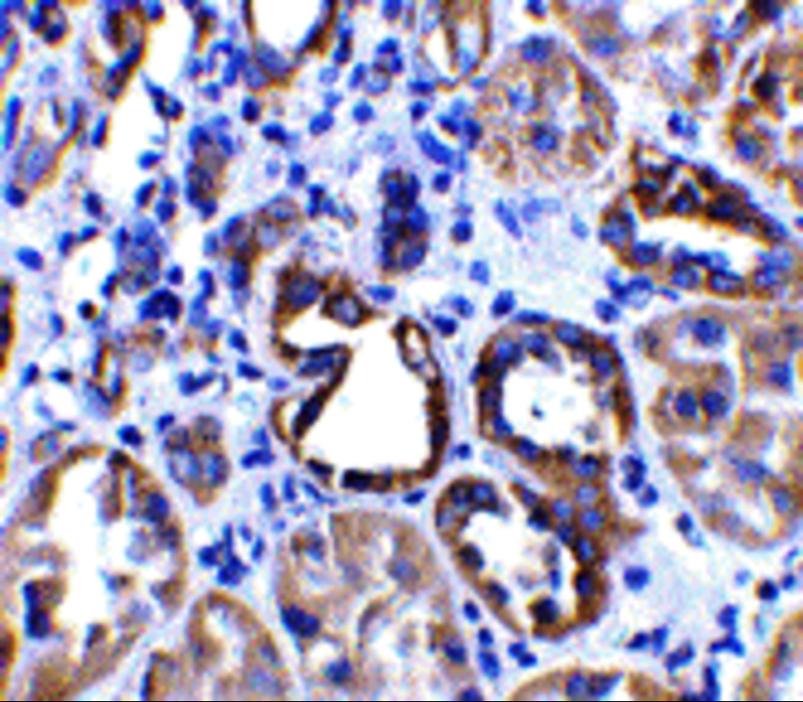
- Application Details WB: 4 µg/mL; IHC: 2 µg/mL; IF: 20 µg/mL. Antibody validated: Western Blot in human, mouse and rat samples; Immunohistochemistry in human, mouse and rat samples; Immunofluorescence in human samples. All other applications and species not yet tested. Biosensis recommends optimal dilutions/concentrations should be determined by the end user.
- Target Angiotensin converting enzyme 2 (ACE2)
- Specificity Anti-ACE2 has no cross response to ACE1. Predicted species reactivity based on immunogen sequence: Bovine: (93%)
- Target Host Species Human
- Species Reactivity Human, Mouse, Rat
- Antibody Host Rabbit
- Antibody Type Polyclonal
- Antibody Isotype IgG
- Conjugate Unconjugated
-
Immunogen Description
ACE2 antibody was raised against a synthetic peptide corresponding to amino acids near the center of human ACE2.
The immunogen is located within amino acids 150 - 200 of ACE2. - Isoform Information Human ACE2 has 2 isoforms, including isoform 1 (805aa, 93kD) and isoform 2 (555aa, 64kD). Mouse ACE2 also has 2 isoforms, including isoform 1 (805aa, 92kD) and isoform 2 (353aa, 40kD). Rat ACE2 has one isoform (805aa, 93kD). 3229 can detect human, mouse and rat.
- Purity Description ACE2 Antibody is affinity chromatography purified via peptide column.
- Format Liquid. ACE2 Antibody is supplied in PBS containing 0.02% sodium azide. Conc.1 mg/mL
- Concentration 1 mg/mL
- Reconstitution Instructions Spin vial briefly before opening.
- Storage Instructions ACE2 antibody can be stored at 2-8°C for three months and -20°C, stable for up to one year. As with all antibodies care should be taken to avoid repeated freeze thaw cycles. Antibodies should not be exposed to prolonged high temperatures.
- Batch Number Please see item label.
- Expiration Date 12 months after date of receipt (unopened vial).
- Alternative Names ACE2 Antibody: ACEH, Angiotensin-converting enzyme 2, ACE-related carboxypeptidase, ACEH, SARS-CoV receptor, SARS-CoV-2 receptor
- Uniprot Number Q9BYF1
- Uniprot Number/Name Q9BYF1 (ACE2_HUMAN)
-
Scientific Background
ACE2 Antibody: Angiotensin-converting enzyme 2 (ACE2) plays a central role in vascular, renal, and myocardial physiology. In contrast to its homolog ACE, ACE2 expression is restricted to heart, kidney, and testis. Recently. ACE2 has also been shown to be a functional receptor of the SARS coronavirus. Homology modeling shows 2019-nCoV has a similar receptor-binding domain structure as SARS-CoV, which suggests COVID-19 (2019-nCoV) may use ACE2 as a receptor in humans for infection. The normal function of ACE2 is to convert the inactive vasoconstrictor angiotensin I (AngI) to Ang1-9 and the active form AngII to Ang1-7, unlike ACE, which converts AngI to AngII. While the role of these vasoactive peptides is not well understood, lack of ACE2 expression in ace2-/ace2- mice leads to severely reduced cardiac contractility, indicating its importance in regulating heart function.
- Research Area Infectious Disease
- Shipping Temperature 2-8°C (on cold packs)
- UNSPSC CODE 41116161
- Regulatory Status For research use only.