SpecificityReacts with ACHE from Human. This antibody is expected to recognise isoform NP_000656.1 only (the ubiquitously expressed, hydrophillic form).
Species ReactivityHuman, Mouse (Predicted), Rat
Immunogen DescriptionA synthetic peptide consisting of amino acids, QFDHYSKQDRCSDL
ConjugateUnconjugated
Purity DescriptionPurified from goat serum by ammonium sulphate precipitation followed by antigen affinity chromatography using the immunizing peptide.
Product DescriptionGoat anti-Acetylcholinesterase (AChE) Polyclonal Antibody (Unconjugated), suitable for Pep-ELISA, WB, ICC, FC.
Application(s)FC, ICC, WB, Pep-ELISA
Application DetailsPeptide ELISA (1:12800), Western Blot (0.1-0.5 µg/mL), Immunofluorescence (10 µg/mL), Flow Cytometry (10 µg/mL). Biosensis recommends optimal dilutions/concentrations should be determined by the end user.
TargetAcetylcholinesterase (AChE)
SpecificityReacts with ACHE from Human. This antibody is expected to recognise isoform NP_000656.1 only (the ubiquitously expressed, hydrophillic form).
Target Host SpeciesHuman
Species ReactivityHuman, Mouse (Predicted), Rat
Antibody HostGoat
Antibody TypePolyclonal
Antibody IsotypeIgG
ConjugateUnconjugated
Immunogen DescriptionA synthetic peptide consisting of amino acids, QFDHYSKQDRCSDL
SequenceQFDHYSKQDRCSDL
Purity DescriptionPurified from goat serum by ammonium sulphate precipitation followed by antigen affinity chromatography using the immunizing peptide.
FormatLiquid antibody. Supplied at 0.5 mg/mL in Tris saline, 0.02% sodium azide, pH 7.3 with 0.5% bovine serum albumin.
Storage InstructionsUpon receipt, aliquot and store at -20°C long-term. Store at 2-8°C short-term (up to 2 weeks). Minimize freezing and thawing.
Batch NumberPlease see item label.
Expiration Date12 months after date of receipt (unopened vial).
Scientific BackgroundTerminates signal transduction at the neuromuscular junction by rapid hydrolysis of the acetylcholine released into the synaptic cleft. Role in neuronal apoptosis (Uniprot).
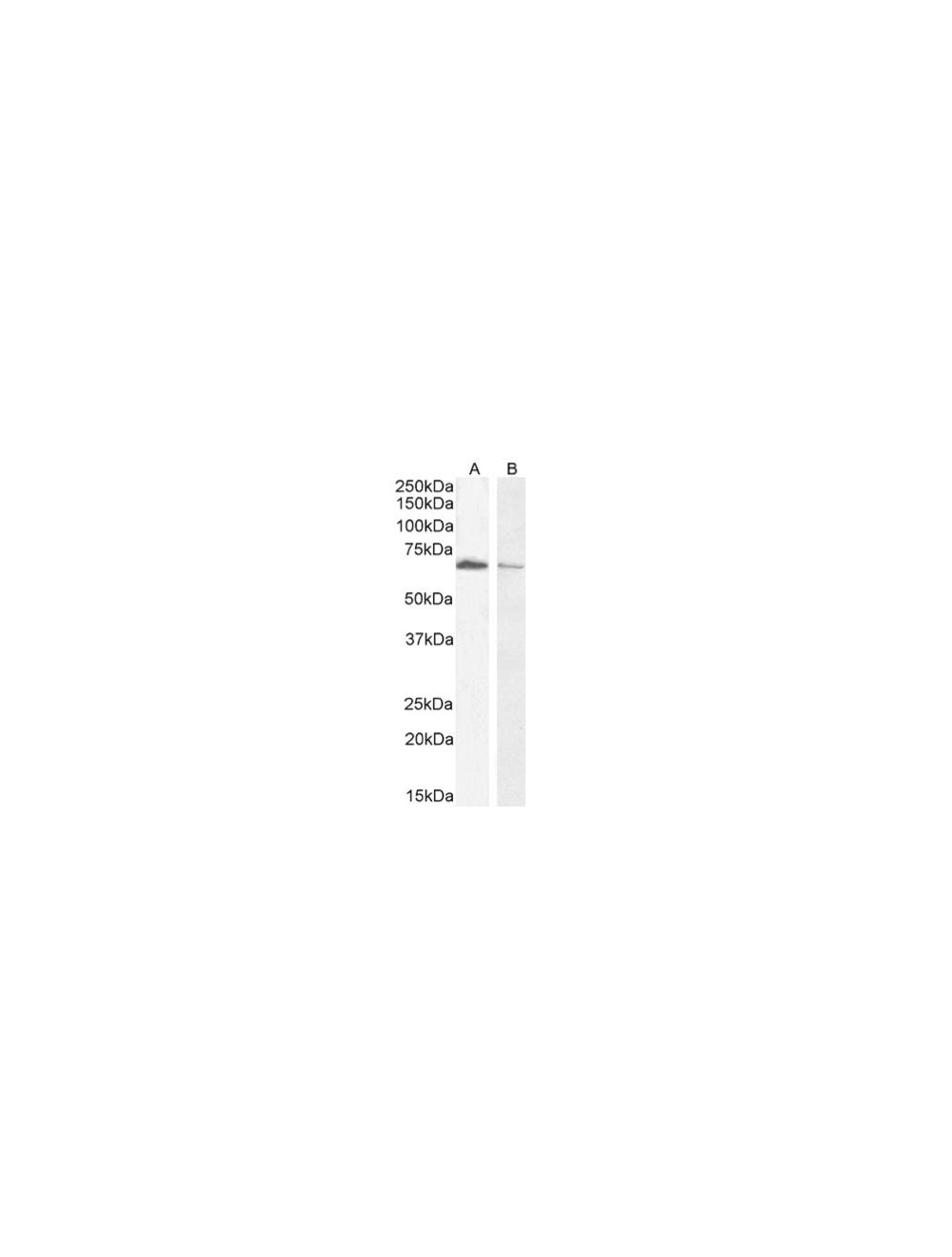
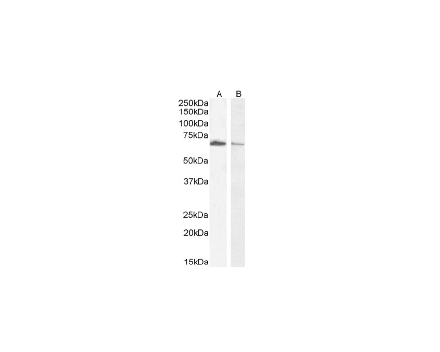
Western Blot staining of AChE in Jurkat (A) and HepG2 (B) cell lysate (35 μg protein in RIPA buffer). Primary antibody concentrations: 0.3 μg/mL (A) and 0.5 μg/mL (B). Detected by chemiluminescence.
A) Immunocytochemistry analysis of paraformaldehyde fixed U2OS cells, permeabilized with 0.15% Triton. Primary incubation 1 hr (10 μg/mL) followed by Alexa Fluor 488 secondary antibody (2 μg/mL). Image shows clear nuclear, membrane and cytoplasmic AChE staining. The nuclear stain is DAPI (blue). Negative control: Unimmunized goat IgG (10 μg/mL), followed by Alexa Fluor 488 secondary antibody (2 μg/mL). B) Flow cytometric analysis of paraformaldehyde fixed HeLa cells (blue line), permeabilized with 0.5% Triton. Primary incubation 1 hr (10 μg/mL) followed by Alexa Fluor 488 secondary antibody (1 μg/mL). IgG control: Unimmunized goat IgG (black line) followed by Alexa Fluor 488 secondary antibody.
 1800 605-5127
 +61 (0)8 8352 7711