SpecificityReacts with Dynactin from Human, Mouse. This antibody is expected to recognise all reported isoforms.
Species ReactivityHuman, Mouse
Immunogen DescriptionA synthetic peptide consisting of amino acids, C-QEQLHQLHSRLIS
ConjugateUnconjugated
Purity DescriptionPurified from goat serum by ammonium sulphate precipitation followed by antigen affinity chromatography using the immunizing peptide.
Application DetailsPeptide ELISA (1:128000), Western Blot (1-2 µg/mL), Flow Cytometry (10 µg/mL). Immunohistochemistry (2-4 µg/mL). Immunofluorescence (10 µg/mL), Flow Cytometry (10 µg/mL). Biosensis recommends optimal dilutions/concentrations should be determined by the end user.
TargetDynactin subunit 1 (DCTN1)
SpecificityReacts with Dynactin from Human, Mouse. This antibody is expected to recognise all reported isoforms.
Target Host SpeciesHuman
Species ReactivityHuman, Mouse
Antibody HostGoat
Antibody TypePolyclonal
Antibody IsotypeIgG
ConjugateUnconjugated
Immunogen DescriptionA synthetic peptide consisting of amino acids, C-QEQLHQLHSRLIS
SequenceQEQLHQLHSRLIS
Purity DescriptionPurified from goat serum by ammonium sulphate precipitation followed by antigen affinity chromatography using the immunizing peptide.
FormatLiquid antibody. Supplied at 0.5 mg/mL in Tris saline, 0.02% sodium azide, pH 7.3 with 0.5% bovine serum albumin.
Storage InstructionsUpon receipt, aliquot and store at -20°C long-term. Store at 2-8°C short-term (up to 2 weeks). Minimize freezing and thawing.
Batch NumberPlease see item label.
Expiration Date12 months after date of receipt (unopened vial).
Scientific BackgroundPlays a key role in dynein-mediated retrograde transport of vesicles and organelles along microtubules by recruiting and tethering dynein to microtubules. Binds to both dynein and microtubules providing a link between specific cargos, microtubules and dynein (Uniprot).
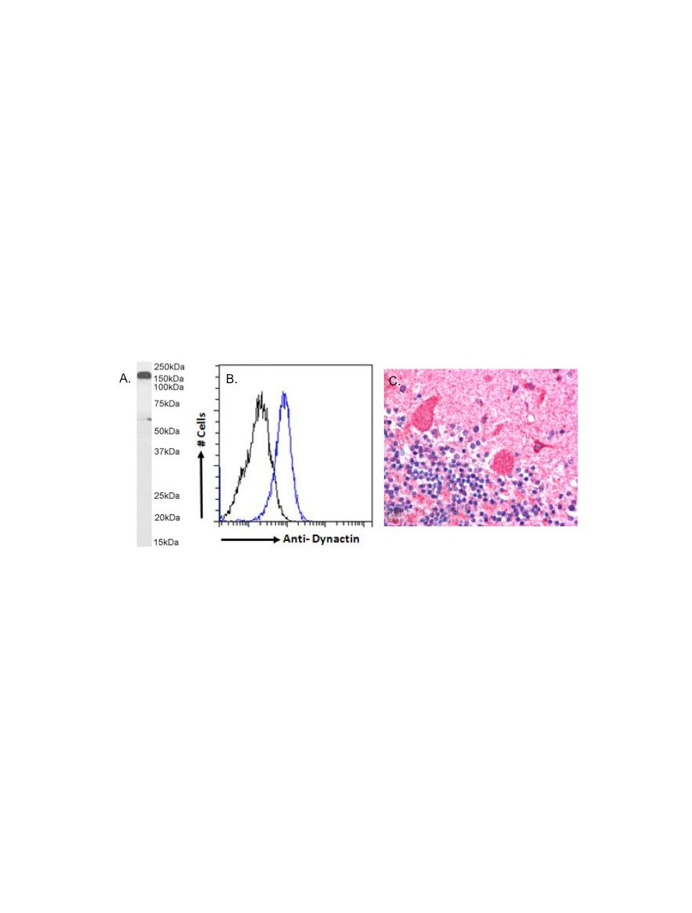
A. Western Blot: (1 µg/mL) staining of Human Cerebellum lysate (35 ug protein in RIPA buffer). Detected by chemiluminescence. B. Flow cytometry analysis: paraformaldehyde fixed HeLa cells (blue line), permeabilized with 0.5% Triton. Primary incubation 1hr (10 µg/mL) followed by Alexa Fluor 488 secondary antibody (1 µg/mL). IgG control: Unimmunized goat IgG (black line) followed by Alexa Fluor 488 secondary antibody. C. Human Cerebellum staining: (2.5 µg/mL) staining of paraffin embedded Human Cerebellum. Steamed antigen retrieval with citrate buffer pH 6, AP-staining.
A. Immunofluorescence analysis: paraformaldehyde fixed HeLa cells, permeabilized with 0.15% Triton. Primary incubation 1hr (10 µg/mL) followed by Alexa Fluor 488 secondary antibody (2 µg/mL), showing cytoplasmic staining. The nuclear stain is DAPI (blue). Negative control: Unimmunized goat IgG (10 µg/mL) followed by Alexa Fluor 488 secondary antibody (2 µg/mL). B. Immunofluorescence analysis: paraformaldehyde fixed U2OS cells, permeabilized with 0.15% Triton. Primary incubation 1hr (10 µg/mL) followed by Alexa Fluor 488 secondary antibody (2 µg/mL), showing cytoplasmic staining. The nuclear stain is DAPI (blue). Negative control: Unimmunized goat IgG (10 µg/mL) followed by Alexa Fluor 488 secondary antibody (2 µg/mL).
 1800 605-5127
 +61 (0)8 8352 7711