 1800 605-5127
 +61 (0)8 8352 7711
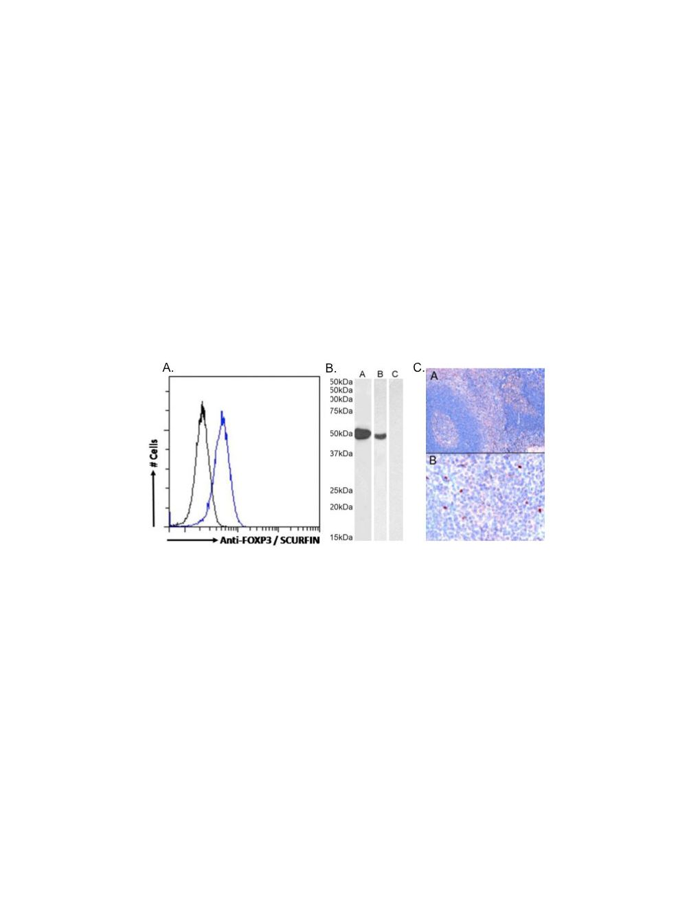

Forkhead box protein P3 (FOXP3), Goat Polyclonal Antibody
- Product Name Forkhead box protein P3 (FOXP3), Goat Polyclonal Antibody
- Product Description Goat anti-Forkhead box protein P3 (FOXP3) Polyclonal Antibody (Unconjugated), suitable for Pep-ELISA, FC.
- Alternative Names FOXP3; forkhead box P3; JM2; AIID; IPEX; PIDX; XPID; DIETER; SCURFIN; scurfin; JM2 protein; immunodeficiency, polyendocrinopathy, enteropathy, X-linked; immune dysregulation, polyendocrinopathy, enteropathy, X-linked
- Application(s) FC, Pep-ELISA
- Antibody Host Goat
- Antibody Type Polyclonal
- Specificity Reacts with FOXP3 from Human.
- Species Reactivity Bovine, Human
- Immunogen Description A synthetic peptide consisting of amino acids, CSNPTPGP
- Conjugate Unconjugated
- Purity Description Purified from goat serum by ammonium sulphate precipitation followed by antigen affinity chromatography using the immunizing peptide.
- Regulatory Status For research use only.
Product Info
- Product Description Goat anti-Forkhead box protein P3 (FOXP3) Polyclonal Antibody (Unconjugated), suitable for Pep-ELISA, FC.
- Application(s) FC, Pep-ELISA
- Application Details ELISA (1:8000), Western Blot (1-3 µg/mL), Immunofluorescence ( 2-4 µg/mL), Flow Cytometry (10 µg/mL). Biosensis recommends optimal dilutions/concentrations should be determined by the end user.
- Target Forkhead box protein P3 (FOXP3)
- Specificity Reacts with FOXP3 from Human.
- Target Host Species Human
- Species Reactivity Bovine, Human
- Antibody Host Goat
- Antibody Type Polyclonal
- Antibody Isotype IgG
- Conjugate Unconjugated
- Immunogen Description A synthetic peptide consisting of amino acids, CSNPTPGP
- Sequence SQRPSRCSNPTPGP
- Purity Description Purified from goat serum by ammonium sulphate precipitation followed by antigen affinity chromatography using the immunizing peptide.
- Format Liquid antibody. Supplied at 0.5 mg/mL in Tris saline, 0.02% sodium azide, pH 7.3 with 0.5% bovine serum albumin.
- Storage Instructions Upon receipt, aliquot and store at -20°C long-term. Store at 2-8°C short-term (up to 2 weeks). Minimize freezing and thawing.
- Batch Number Please see item label.
- Expiration Date 12 months after date of receipt (unopened vial).
- Alternative Names FOXP3; forkhead box P3; JM2; AIID; IPEX; PIDX; XPID; DIETER; SCURFIN; scurfin; JM2 protein; immunodeficiency, polyendocrinopathy, enteropathy, X-linked; immune dysregulation, polyendocrinopathy, enteropathy, X-linked
- Uniprot Number Q9BZS1
- Uniprot Number/Name Q9BZS1 (FOXP3_HUMAN)
- Scientific Background Transcriptional regulator which is crucial for the development and inhibitory function of regulatory T-cells (Treg). Plays an essential role in maintaining homeostasis of the immune system by allowing the acquisition of full suppressive function and stability of the Treg lineage, and by directly modulating the expansion and function of conventional T-cells.
- Shipping Temperature 2-8°C (on cold packs)
- UNSPSC CODE 41116161
- Regulatory Status For research use only.


