Product DescriptiongoogleGoat anti-Histone deacetylase 2 (HD2) Polyclonal Antibody (Unconjugated), suitable for Pep-ELISA, WB, ICC, FC.
Alternative NamesHdac2; histone deacetylase 2; D10Wsu179e; YAF1; Yy1bp; mRPD3; HD2; OTTMUSP00000022803; YY1 transcription factor-binding protein
Application(s)FC, ICC, WB, Pep-ELISA
Antibody HostGoat
Antibody TypePolyclonal
SpecificityReacts with Hdac2 (mouse) from Human. This antibody imay cross-react with HDAC1.
Species ReactivityBovine (Predicted), Dog (Predicted), Human, Mouse, Pig (Predicted), Rat (Predicted)
Immunogen DescriptionA synthetic peptide consisting of amino acids, C-PEDAVHEDSGDE
ConjugateUnconjugated
Purity DescriptionPurified from goat serum by ammonium sulphate precipitation followed by antigen affinity chromatography using the immunizing peptide.
Product DescriptionGoat anti-Histone deacetylase 2 (HD2) Polyclonal Antibody (Unconjugated), suitable for Pep-ELISA, WB, ICC, FC.
Application(s)FC, ICC, WB, Pep-ELISA
Application DetailsPeptide ELISA (1:2000), Western Blot (1-2 µg/mL), Immunocytochemistry (10 µg/mL),Flow Cytometry (10 µg/mL). Biosensis recommends optimal dilutions/concentrations should be determined by the end user.
TargetHistone deacetylase 2 (HD2)
SpecificityReacts with Hdac2 (mouse) from Human. This antibody imay cross-react with HDAC1.
Target Host SpeciesHuman
Species ReactivityBovine (Predicted), Dog (Predicted), Human, Mouse, Pig (Predicted), Rat (Predicted)
Antibody HostGoat
Antibody TypePolyclonal
Antibody IsotypeIgG
ConjugateUnconjugated
Immunogen DescriptionA synthetic peptide consisting of amino acids, C-PEDAVHEDSGDE
SequencePEDAVHEDSGDE
Purity DescriptionPurified from goat serum by ammonium sulphate precipitation followed by antigen affinity chromatography using the immunizing peptide.
FormatLiquid antibody. Supplied at 0.5 mg/mL in Tris saline, 0.02% sodium azide, pH 7.3 with 0.5% bovine serum albumin.
Storage InstructionsUpon receipt, aliquot and store at -20°C long-term. Store at 2-8°C short-term (up to 2 weeks). Minimize freezing and thawing.
Batch NumberPlease see item label.
Expiration Date12 months after date of receipt (unopened vial).
Alternative NamesHdac2; histone deacetylase 2; D10Wsu179e; YAF1; Yy1bp; mRPD3; HD2; OTTMUSP00000022803; YY1 transcription factor-binding protein
Scientific BackgroundResponsible for the deacetylation of lysine residues on the N-terminal part of the core histones (H2A, H2B, H3 and H4). Histone deacetylation gives a tag for epigenetic repression and plays an important role in transcriptional regulation, cell cycle progression and developmental events. Histone deacetylases act via the formation of large multiprotein complexes. Forms transcriptional repressor complexes by associating with MAD, SIN3, YY1 and N-COR.
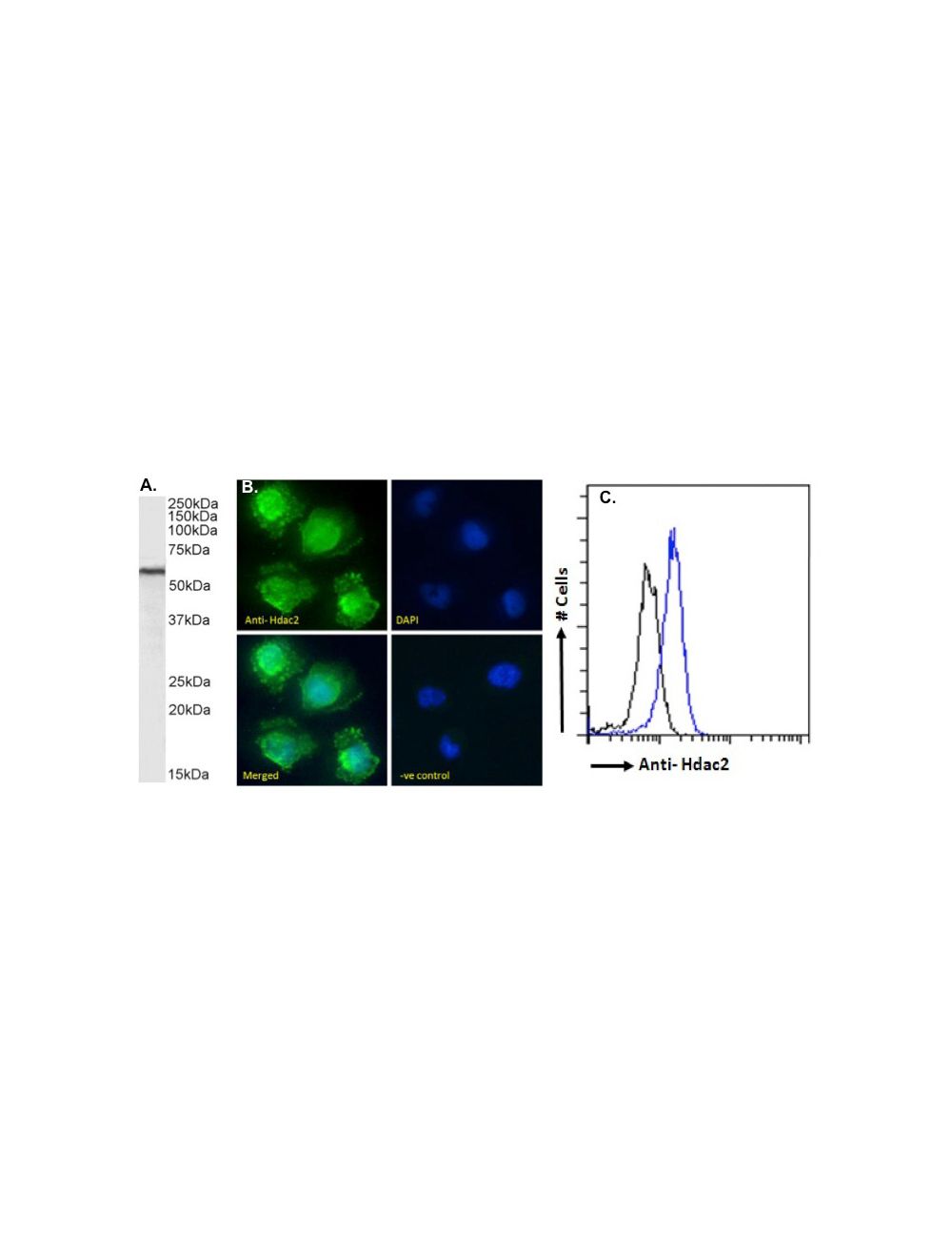
A. Western Blot: (1 µg/mL) staining of HEK293 nuclear cell lysate. (35 ug protein in RIPA buffer). Detected by Chemiluminescence B. Immunofluorescenceanalysis: of paraformaldehyde fixed U251 cells, permeabilized with 0.15% Triton. Primary incubation 1hr (10 µg/mL) followed by Alexa Fluor 488 secondary antibody (2 µg/mL), showing nuclear staining. The nuclear stain is DAPI (blue). Negative control: Unimmunized goat IgG (10 µg/mL) followed by Alexa Fluor 488 secondary antibody (2 µg/mL). C. Flow cytometryanalysis: of paraformaldehyde fixed HeLa cells (blue line), permeabilized with 0.5% Triton. Primary incubation 1hr (10 µg/mL) followed by Alexa Fluor 488 secondary antibody (2 µg/mL). IgG control: Unimmunized goat IgG (black line) followed by Alexa Fluor 488 secondary antibody.
G-1926-100 Immunocytochemistry analysis of paraformaldehyde fixed NIH3T3 cells, permeabilized with 0.15% Triton. Primary incubation 1hr (10 μg/mL) followed by Alexa Fluor 488 secondary antibody (2 μg/mL), showing membrane, cytoplasmic and nuclear staining. The nuclear stain is DAPI (blue). Negative control: Unimmunized goat IgG (10 μg/mL) followed by Alexa Fluor 488 secondary antibody (2 μg/mL).
 1800 605-5127
 +61 (0)8 8352 7711