SpecificityReacts with KIT from Human, Mouse. This antibody is expected to recognise both reported isoforms (NP_000213.1; NP_001087241.1).
Species ReactivityHuman, Mouse
Immunogen DescriptionA synthetic peptide consisting of amino acids, C-HHGDFNYERQAT
ConjugateUnconjugated
Purity DescriptionPurified from goat serum by ammonium sulphate precipitation followed by antigen affinity chromatography using the immunizing peptide.
Product DescriptionGoat anti-Mast/stem cell growth factor receptor Kit (SCFR) Polyclonal Antibody (Unconjugated), suitable for Pep-ELISA, ICC, IHC, FC.
Application(s)FC, ICC, IHC, Pep-ELISA
Application DetailsPeptide ELISA (1:16,000), Immunohistochemistry (5 µg/mL), Immunocytochemistry (10 µg/mL), Flow Cytometry (10 µg/mL). Biosensis recommends optimal dilutions/concentrations should be determined by the end user.
SpecificityReacts with KIT from Human, Mouse. This antibody is expected to recognise both reported isoforms (NP_000213.1; NP_001087241.1).
Target Host SpeciesHuman
Species ReactivityHuman, Mouse
Antibody HostGoat
Antibody TypePolyclonal
Antibody IsotypeIgG
ConjugateUnconjugated
Immunogen DescriptionA synthetic peptide consisting of amino acids, C-HHGDFNYERQAT
SequenceHHGDFNYERQAT
Purity DescriptionPurified from goat serum by ammonium sulphate precipitation followed by antigen affinity chromatography using the immunizing peptide.
FormatLiquid antibody. Supplied at 0.5 mg/mL in Tris saline, 0.02% sodium azide, pH 7.3 with 0.5% bovine serum albumin.
Storage InstructionsUpon receipt, aliquot and store at -20°C long-term. Store at 2-8°C short-term (up to 2 weeks). Minimize freezing and thawing.
Batch NumberPlease see item label.
Expiration Date12 months after date of receipt (unopened vial).
Scientific BackgroundTyrosine-protein kinase that acts as cell-surface receptor for the cytokine KITLG/SCF and plays an essential role in the regulation of cell survival and proliferation, hematopoiesis, stem cell maintenance, gametogenesis, mast cell development, migration and function, and in melanogenesis.
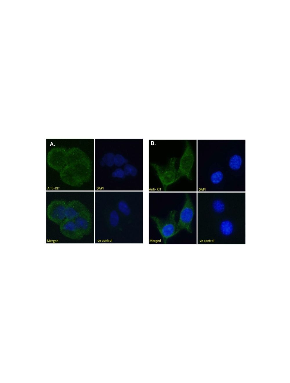
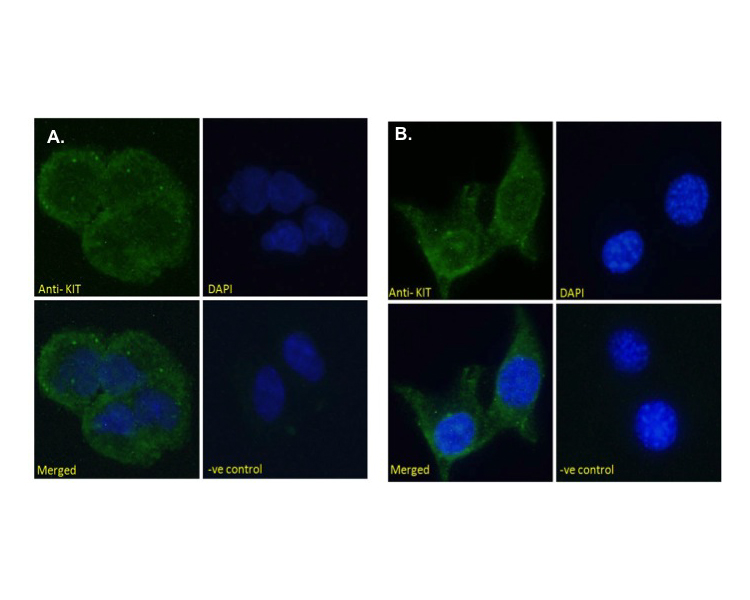
A. Immunofluorescence analysis: of paraformaldehyde fixed HEK293 cells, permeabilized with 0.15% Triton. Primary incubation 1hr (10 µg/mL) followed by Alexa Fluor 488 secondary antibody (2 µg/mL), showing cytoplasmic staining. The nuclear stain is DAPI (blue). Negative control: Unimmunized goat IgG (10 µg/mL) followed by Alexa Fluor 488 secondary antibody (2 µg/mL). B. Immunofluorescenceanalysis: of paraformaldehyde fixed NIH3T3 cells, permeabilized with 0.15% Triton. Primary incubation 1hr (10 µg/mL) followed by Alexa Fluor 488 secondary antibody (2 µg/mL), showing membrane staining. The nuclear stain is DAPI (blue). Negative control: Unimmunized goat IgG (10 µg/mL) followed by Alexa Fluor 488 secondary antibody (2 µg/mL).
A. Flow cytometric analysis: of paraformaldehyde fixed MCF7 cells (blue line), permeabilized with 0.5% Triton. Primary incubation 1hr (10 µg/mL) followed by Alexa Fluor 488 secondary antibody (1 µg/mL). IgG control: Unimmunized goat IgG (black line) followed by Alexa Fluor 488 secondary antibody. B. Staining of Human Skin: (5 µg/mL) staining of paraffin embedded Human Skin. Steamed antigen retrieval with citrate buffer pH 6, AP-staining.
 1800 605-5127
 +61 (0)8 8352 7711