 1800 605-5127
 +61 (0)8 8352 7711
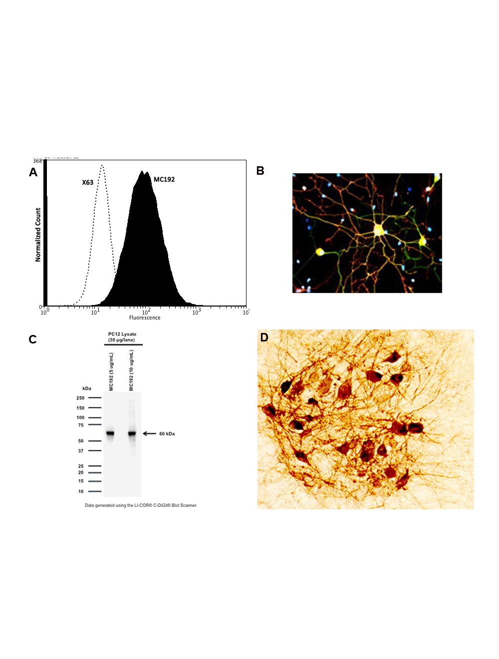

NGFR/p75 neurotrophin receptor (p75NTR), Mouse Monoclonal Antibody
- Product Name NGFR/p75 neurotrophin receptor (p75NTR), Mouse Monoclonal Antibody
- Product Description Mouse anti-p75 neurotrophin receptor (p75NTR) Monoclonal Antibody (Unconjugated), suitable for WB, IHC-Frozen, ELISA.
- Alternative Names Low-affinity nerve growth factor receptor; NGF receptor; Gp80-LNGFR; p75 ICD; Low affinity neurotrophin receptor p75NTR; CD271
- Application(s) ELISA, IHC-Frozen, WB
- Antibody Host Mouse
- Antibody Type Monoclonal
- Specificity MC192 is specific only for RAT NGFR, no reactivity to Human or Mouse NGFR has been reported This monoclonal antibody has been tested for immunohistochemical localisation of p75NTR-expressing rat cells in the spinal cord and brain. This monoclonal antibody does not cross react with p75NTR-expressing cells in other species.
- Species Reactivity Rat
- Immunogen Description NGF receptor
- Conjugate Unconjugated
- Purity Description Protein G purified immunoglobulin
- Regulatory Status For research use only.
Product Info
- Product Description Mouse anti-p75 neurotrophin receptor (p75NTR) Monoclonal Antibody (Unconjugated), suitable for WB, IHC-Frozen, ELISA.
- Application(s) ELISA, IHC-Frozen, WB
- Application Details IH (lightly fixed), ELISA, WB, Flow Cytometry (2 ug per 10^6 cells) IP (non-reducing conditions only!; do not use reducing agents such as DTT or beta-mercaptoethanol), Traditional formalin fixed paraffin embedded immunohistochemistry is NOT recommended with MC192. Motor neuron isolation, Gene/Toxin Delivery to rat sensory/motor neurons. A working solution of 1-2 µg/mL was determined by immunohistochemical staining on 4% paraformaldehyde fixed, or alcohol fixed rat spinal cord and brain. For non-denatured WB, 1-5 µg/mL was found to be suitable with suitable controls (PC12 lysate). ELISA: detection only, 1-5 µg/mL has been suggested in literature.Immunoprecipitation: 5 µg/mL, > 0.5% triton X-100 buffer/500 ug/lysate; PC12 positive control strong suggested. MC192 is not suitable as a blocking agent, although it has been incorrectly used for this purpose in many published works. The antibody was generated specifically by screening for monoclonals that had the ability to ENHANCE the binding of NGF, the natural ligand for p75. Therefore, this antibody is particularly unusual. The full details can be found in the original paper, which is listed on our datasheet (see Chandler et al, 1984). Biosensis recommends optimal dilutions/concentrations should be determined by the end user.
- Target NGFR/p75 neurotrophin receptor (p75NTR)
- Specificity MC192 is specific only for RAT NGFR, no reactivity to Human or Mouse NGFR has been reported This monoclonal antibody has been tested for immunohistochemical localisation of p75NTR-expressing rat cells in the spinal cord and brain. This monoclonal antibody does not cross react with p75NTR-expressing cells in other species.
- Target Host Species Rat
- Species Reactivity Rat
- Antibody Host Mouse
- Antibody Type Monoclonal
- Antibody Isotype IgG1
- Clone Name MC192
- Conjugate Unconjugated
- Immunogen Description NGF receptor
- Purity Description Protein G purified immunoglobulin
- Format Lyophilized
- Reconstitution Instructions Spin vial briefly before opening. Reconstitute in 100 µL sterile-filtered, ultrapure water. Centrifuge to remove any insoluble material.
- Storage Instructions The MC192 is supplied in lyophilized form from Protein G-purified hybridoma cell culture supernatants. The lyophilized antibody is stable when stored at 2-8°C or -20°C. After reconstitution undiluted aliquots should be kept at -20°C for up to six months. For additional stability Glycerol (1:1) may be added after reconstitution. Repetitive freeze/thaw cycle should be avoided.
- Batch Number Please see item label.
- Expiration Date 12 months after date of receipt (unopened vial).
- Alternative Names Low-affinity nerve growth factor receptor; NGF receptor; Gp80-LNGFR; p75 ICD; Low affinity neurotrophin receptor p75NTR; CD271
- Uniprot Number P07174
- Uniprot Number/Name P07174 (TNR16_RAT)
- Scientific Background Monoclonal antibody MC192 against the rat low affinity nerve growth factor receptor (p75NTR) is derived from the fusion of Sp2/0-Ag 14 myeloma cells with mouse immune splenocytes. MC192 monoclonal antibody was originally generated by Chandlers et al. p75NTR was originally discovered as a low affinity nerve growth factor receptor. Later it was found that it was the receptor for all neurotrophins. It mediates signals of neurotrophins for neuronal survival, apoptosis, neurite outgrowth and synaptic plasticity. Recently, it has been revealed that p75NTR not only acts as the receptor for neurotrophins but also the receptor for many other pathological ligands such as prions, rabies virus and amyloid beta. p75NTR also acts as a co-receptor for NOGO which mediates inhibitory signals of myelin associated protein. p75NTR is highly expressed in a number of non-neuronal and neuronal cells including motor neurons during development and also in damaged neurons. MC192 recognizes the extracellular domain of the neurotrophin receptor p75NTR in rat. MC192 antibody may be used for immunocytochemical localisation of rat cells expressing p75NTR, ELISA and western blot. This antibody has also been used for the construction of the MC192-saporin immunotoxin for specific elimination of neuronal populations in basal forebrain cholinergic neurons to generate an animal model for Alzheimer's disease. Using Flow Cytometry, this antibody has frequently been employed for panning to isolate p75NTR-expressing rat cells. MC192 has a potential use as the ligand for gene delivery into p75NTR-expressing rat cells via a receptor-mediated mechanism. FUNCTION: Low affinity receptor which can bind to NGF, BDNF, NT-3, and NT-4. Can mediate cell survival as well as cell death of neural cells. SUBUNIT: Homodimer; disulfide-linked. Interacts with p75NTR-associated cell death executor. Interacts with NGFRAP1/BEX3. Interacts with TRAF2, TRAF4, TRAF6, PTPN13 and RANBP9. Interacts through TRAF6 with SQSTM1 which bridges NGFR to NTRK1 (By similarity). Interacts with BEX1. SUBCELLULAR LOCATION: Membrane; single-pass type I membrane protein. DOMAIN: Death domain is responsible for interaction with RANBP9. PTM: N- and O-glycosylated. PTM: Phosphorylated on serine residues. SIMILARITY: Contains 1 death domain. SIMILARITY: Contains 4 TNFR-Cys repeats.
- Shipping Temperature 25°C (ambient)
- UNSPSC CODE 41116161
- Regulatory Status For research use only.
Specifications
-
Specific References
Riffault B, Kourdougli N, Dumon C, Ferrand N, Buhler E, Schaller F, Chambon C, Rivera C, Gaiarsa JL, Porcher C (2016) Pro-Brain-Derived Neurotrophic Factor (proBDNF)-Mediated p75NTR Activation Promotes Depolarizing Actions of GABA and Increases Susceptibility to Epileptic Seizures. Cereb. Cortex [Epub ahead of print]. Application: Western Blot; Species: Rat
Brandli A, Johnstone DM, Stone J (2016) Remote Ischemic Preconditioning Protects Retinal Photoreceptors: Evidence From a Rat Model of Light-Induced Photoreceptor Degeneration. Invest Ophthalmol Vis Sci. 57(13):5302-13 Application: Western Blot, IHC; Species: Rat
Riffault B, Medina I, Dumon C, Thalman C, Ferrand N, Friedel P, Gaiarsa JL, Porcher C. (2014) "Pro-Brain-Derived Neurotrophic Factor Inhibits GABAergic Neurotransmission by Activating Endocytosis and Repression of GABAA Receptors." J. Neurosci. 34(40):13516-34 Application: Western Blot,Neuronal cells and hippocampi; Species: Rat
Kalincik T et al (2011) Selected changes in spinal cord morphology after T4 transection and olfactory ensheathing cell transplantation. Auton Neurosci. 158(1-2):31-8 Application: IF; Species: Rat
Wu A et al (2011) Delayed olfactory ensheathing cell transplants reduce nociception after dorsal root injury. Exp Neurol. 229(1):143-57 Application: IF; Species: Rat
Davies A et al (2010) The alpha2delta subunits of voltage-gated calcium channels form GPI-anchored proteins, a post translational modification essential for function Proc Natl Acad Sci U S A. Jan 26;107(4):1654-9
Kalincik T et al (2010) Olfactory ensheathing cells reduce duration of autonomic dysreflexia in rats with high spinal cord injury. Auton Neurosci. 154 (1-2):20-9 Application: IHC; Species: Rat
Wilson-Gerwing T.D. et al (2009) J Comp Neurol. 2009 Sep 1;516(1):49-58
Feron F et al (2008) Neurotrophin expression in the adult olfactory epithelium. Brain Res. 1196:13-21 Application: IHC; Species: Rat
Bianco JI et al (2004) Neurotrophin 3 promotes purification and proliferation of olfactory ensheathing cells from human nose. Glia. 45(2):111-23 Application: IHC, IF; Species: Rat
Eyles D et al (2003) Neuroscience. 2003;118(3):641-53. Application: IHC; Species: Rat
Lu J et al (2001) Transplantation of nasal olfactory tissue promotes partial recovery in paraplegic adult rats. Brain Res. 889(1-2):344-57 Application: IF; Species: Rat -
General References
Vilar M. et al (2009) Activation of the p75 neurotrophin receptor through conformational rearrangement of disulphide-linked receptor dimers Neuron. 2009 Apr 16;62(1):72-83
Luther, J.A. and Birren, S.J. (2009) J. Neurosci 29, 5411-24
Lagares, A. et al. (2007) J. Neurosci 27, 7939-53
Kruger, G. M. et al. (2002) Neuron 35, 657-69
Tuffereau, C. et al (1998) Embo J 17, 7250-9
Zhou, X. F., and Rush, R. A. (1996) J Comp Neurol 372, 37-48
Huber, J., Dittrich, F., and Phelan, P. (1993) Eur J Biochem 218, 1031-1039
Rabizadeh S. et al (1993) Science. Jul 16;261(5119):345-8.
Stemple, D. L. & Anderson, D. J. (1992) Cell 71, 973-85
Urschel, B. A. & Hulsebosch, C. E. (1992) Brain Res Dev Brain Res 69, 261-70
Birren SJ et al (1992) Science. Jul 17;257(5068):395-7.
Yan, Q., and Johnson, E. M., Jr. (1988) J Neurosci. 1988 Sep;8(9):3481-98.
Johnson, D. et al. (1986) Cell 47, 545-54
Chandler, C. E. et al (1984) (1984) J Biol Chem 259, 6882-6889