Immunofluorescence analysis of TrkA expression on human SHSY-5Y neuroblastoma cell membrane. Fixed (4% formaldehyde), non-permeabilized, and blocked (10% normal horse serum) SHSY-5Y cells were incubated with TrkA antibody M-1723-100 (extracellular domain, 2 µg/mL, green) for 1 hour. Primary antibody binding was visualized with a secondary donkey anti-mouse-CF488A antibody (4 µg/mL, 1 hour incubation). Cell nuclei were stained with Hoechst dye (blue). Magnification: 100x.
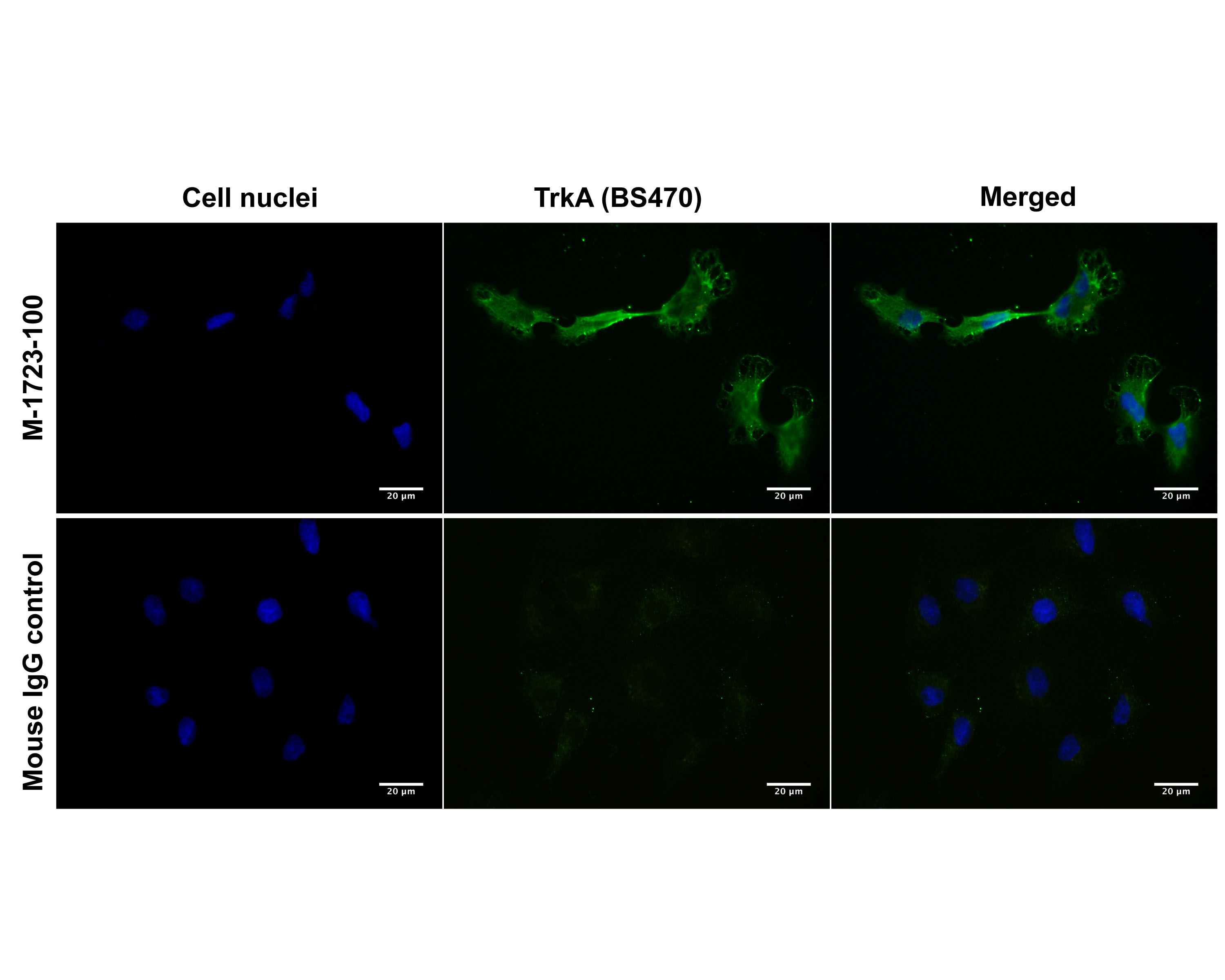
Immunofluorescence analysis of TrkA expression in human SHSY-5Y neuroblastoma cells, comparing TrkA antibodies M-1723-100 (BS470, extracellular domain) and M-1719-100 (BS292, intracellular domain). Cells were fixed (4% formaldehyde, 10 minutes), permeabilized (0.1% Triton X100, A and C) or non-permeabilized (A), and blocked (10% normal horse serum) for 30 minutes. SHSY-5Y cells were incubated for 1 hour with TrkA primary antibodies (2 µg/mL, green) M-1723-100 (extracellular domain, A1 and B1) and M-1719-100 (intracellular domain, C1). Primary antibody binding was visualized with a secondary donkey anti-mouse-CF488A antibody (4 µg/mL, 1 hour incubation). Cell nuclei were stained with Hoechst dye (blue). Control cells were treated exactly the same way, substituting the primary TrkA antibody with normal mouse IgG (A2, B2 and C2) and using the same camera exposure times to record images. Magnification: 100x. This batch of SHSY-5Y cells did not show TrkA expression on the cell membrane (A), while both antibodies were immunoreactive for intracellulary stored TrkA receptor (B and C).
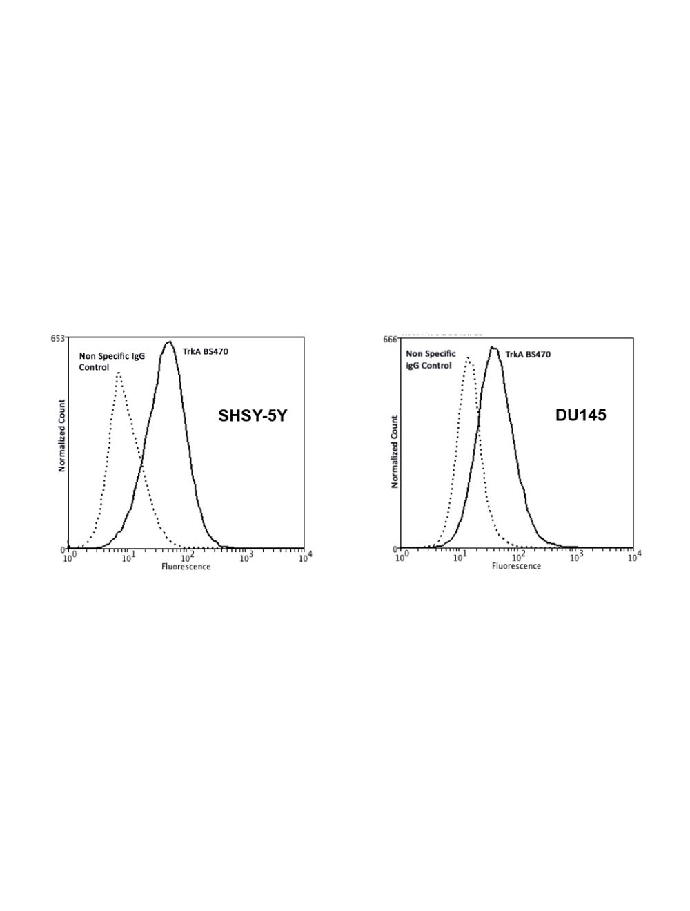
Comparison of TrkA expression on non-permeabilized and permeabilized human SHSY-5Y neuroblastoma cells by Flow Cytometry. This batch of SHSY-5Y cells did not show TrkA expression on the membrane (left image), but immunoreactivity in permeabilized cells (right image), suggesting that TrkA is stored intracellularly. Conditions: Permeabilization with absolute methanol (10 minutes on ice), or no permeabilization (incubation of primary antibody on ice only). Blocking: 200 µg/mL normal sheep IgG (30 minutes) on ice. Primary antibody: Mouse Monoclonal antibody to TrkA, extracellular domain (cat# M-1723-100, 2 µg per ~106 cells) for 30 minutes on ice. Secondary antibody: Goat anti-mouse PE (1:100 dilution, 20 min in dark on ice. Non-specific Control IgG, clone X63 (cat# M-1249-100) was used as negative control under same conditions (black dashed). Flow cytometry data and results were generated using Orflo MoxiflowTM instrument and protocols.
 1800 605-5127
 +61 (0)8 8352 7711