 1800 605-5127
 +61 (0)8 8352 7711
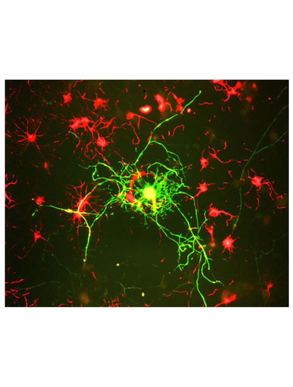

Neurofilament medium polypeptide (NF-M), Mouse Monoclonal Antibody (3H11)
- Product Name Neurofilament medium polypeptide (NF-M), Mouse Monoclonal Antibody (3H11)
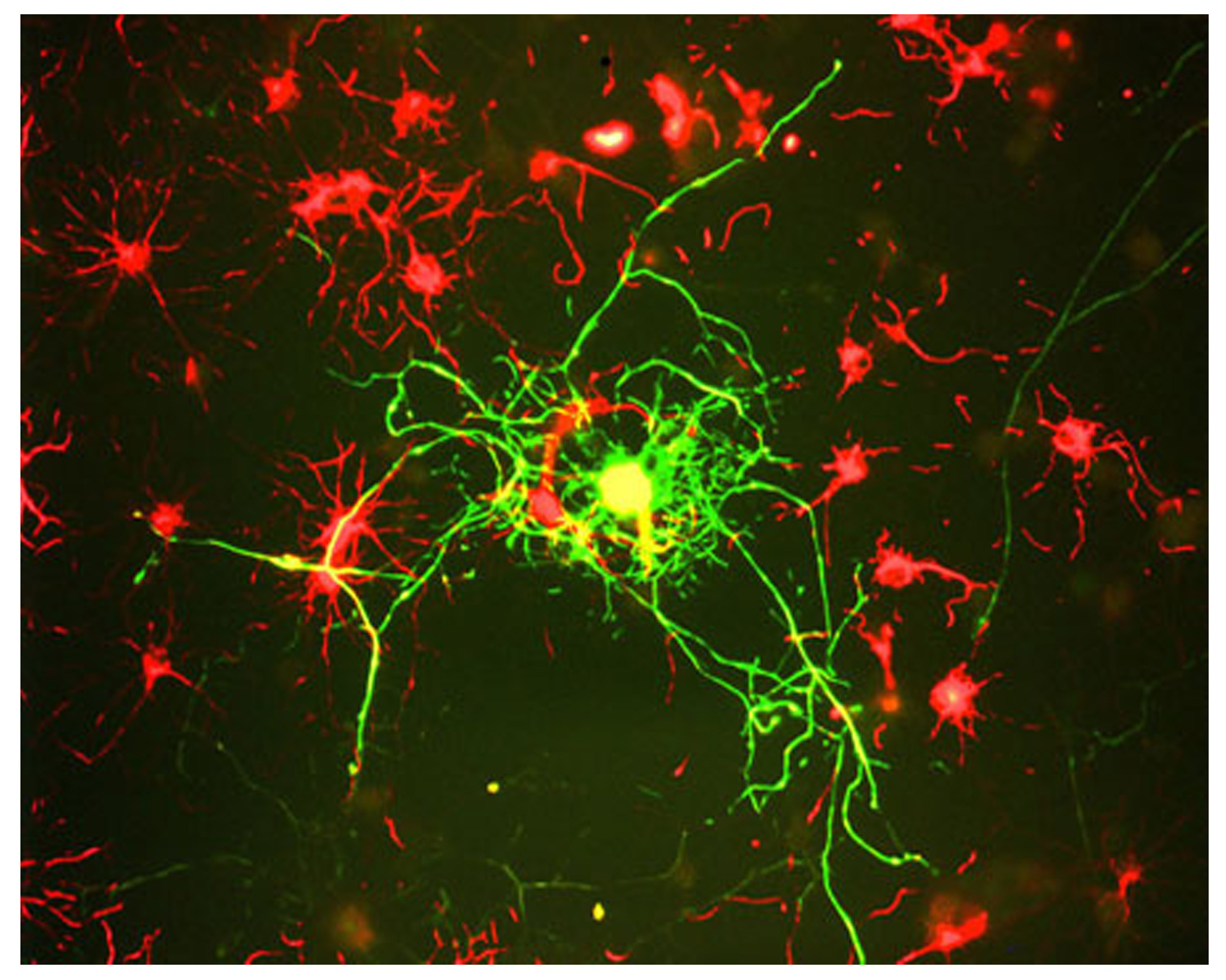
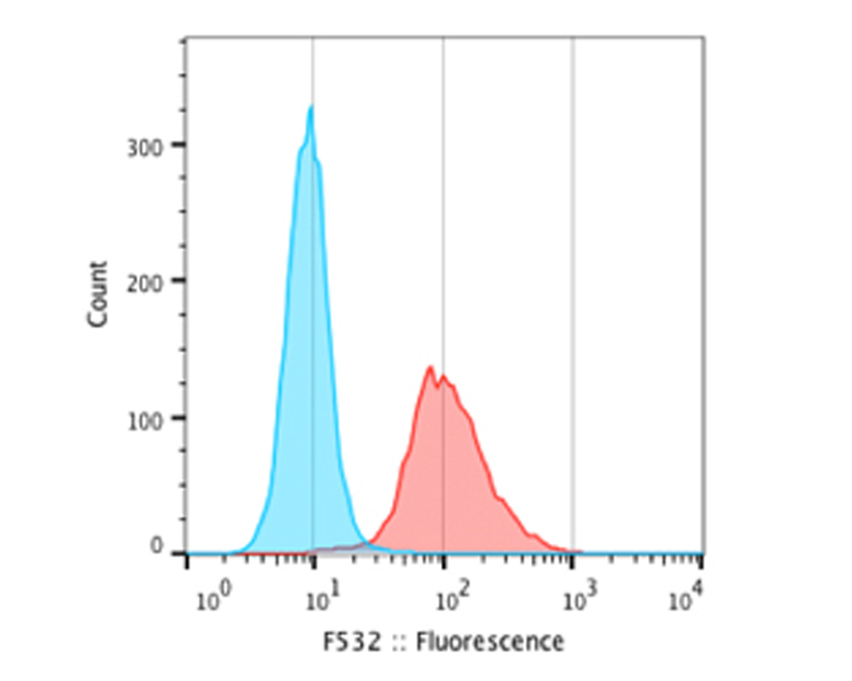
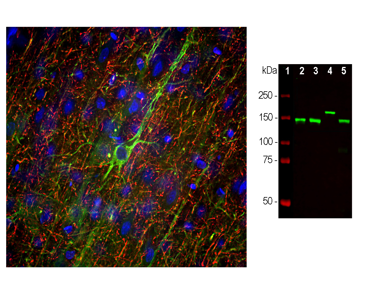
- Product Description Mouse anti-Neurofilament medium polypeptide (NF-M), Monoclonal Antibody (Unconjugated), suitable for WB, Immunostaining and FC.
- Alternative Names NF-M; NFM; Neurofilament medium polypeptide; 160 kDa neurofilament protein; Neurofilament 3; Neurofilament triplet M protein; Nefm; Nef3; Nfm
- Application(s) FC, IF, ICC, IHC, WB
- Antibody Host Mouse
- Antibody Type Monoclonal
- Specificity Species cross-reactivity includes human, rat, mouse, cow, pig, horse and chicken.
- Species Reactivity Bovine, Chicken, Horse, Human, Mouse, Pig, Rat
- Immunogen Description This antibody has been made against a recombinant fusion protein containing the extreme C-terminus of rat NF-M (amino acids 677-845) expressed in and purified from E. coli. The epitope is localized to within the last 56 amino acids at the extreme C-terminus of rat NF-M, the so-called KE segment which is highly conserved between NF-M molecules from different species.
- Conjugate Unconjugated
- Purity Description Protein G purified
- Regulatory Status For research use only.
Product Info
- Product Description Mouse anti-Neurofilament medium polypeptide (NF-M), Monoclonal Antibody (Unconjugated), suitable for WB, Immunostaining and FC.
-
Related Products
Neurofilament medium polypeptide (NF-M), Chicken Polyclonal Antibody
Neurofilament medium polypeptide (NF-M), Rabbit Polyclonal Antibody
- Application(s) FC, IF, ICC, IHC, WB
- Application Details Western blot (WB), Immunocytochemistry (ICC) / Immunofluorescence (IF), Immunohistochemistry (IHC) and Flow Cytometry (FC). A dilution of 1:5,000 is recommended for WB. A dilution of 1:2,000 is recommended for ICC/IFF and IHC. A dilution of 2 ug per 10^6 cells is recommended for FC. Biosensis recommends optimal dilutions/concentrations should be determined by the end user.
- Target Neurofilament medium polypeptide (NF-M)
- Specificity Species cross-reactivity includes human, rat, mouse, cow, pig, horse and chicken.
- Target Host Species Rat
- Species Reactivity Bovine, Chicken, Horse, Human, Mouse, Pig, Rat
- Antibody Host Mouse
- Antibody Type Monoclonal
- Antibody Isotype IgG1
- Clone Name 3H11
- Conjugate Unconjugated
- Immunogen Description This antibody has been made against a recombinant fusion protein containing the extreme C-terminus of rat NF-M (amino acids 677-845) expressed in and purified from E. coli. The epitope is localized to within the last 56 amino acids at the extreme C-terminus of rat NF-M, the so-called KE segment which is highly conserved between NF-M molecules from different species.
- Purity Description Protein G purified
- Format Lyophilized from PBS buffer pH 7.2-7.6 with 0.1% trehalose, and sodium azide
- Reconstitution Instructions Spin vial briefly before opening. Reconstitute with 100 µL sterile-filtered, ultrapure water to achieve a 1 mg/mL concentration. Centrifuge to remove any insoluble material.
- Storage Instructions Store lyophilized antibody at 2-8°C After reconstitution of lyophilized antibody, aliquot and store at -20°C for a higher stability. Avoid freeze-thaw cycles. Store at 4°C for up to one month for short term storage and frequent use.
- Batch Number Please see item label.
- Expiration Date 12 months after date of receipt (unopened vial).
- Alternative Names NF-M; NFM; Neurofilament medium polypeptide; 160 kDa neurofilament protein; Neurofilament 3; Neurofilament triplet M protein; Nefm; Nef3; Nfm
- Uniprot Number P07197
- Uniprot Number/Name P07197 (NFM_HUMAN)
- Scientific Background Neurofilaments are the 10nm or intermediate filament proteins found specifically in neurons, and are composed predominantly of three major proteins called NF-L, NF-M and NF-H, though other filament proteins may be included also. The major function of neurofilaments is likely to control the diameter of large axons. NF-L is the neurofilament light or low molecular weight polypeptide and runs on SDS-PAGE gels at 68-70kDa with some variability across species. Antibodies to NF-L are useful for identifying neuronal cells and their processes in cell culture and sectioned material. NF-L antibody can also be useful for the visualization of neurofilament rich accumulations seen in many neurological diseases, such as Lou Gehrig’s disease (ALS), giant axon neuropathy, Charcot-Marie Tooth disease and others. (Ref: uniprot.org)
- Shipping Temperature 25°C (ambient)
- UNSPSC CODE 41116161
- Regulatory Status For research use only.
Specifications
-
Specific References
Felitsyn N. et al (2008) The heme precursor delta-aminolevulinate blocks peripheral myelin formation. J Neurochem. 2008 Sep;106(5):2068-79.