 1800 605-5127
 +61 (0)8 8352 7711
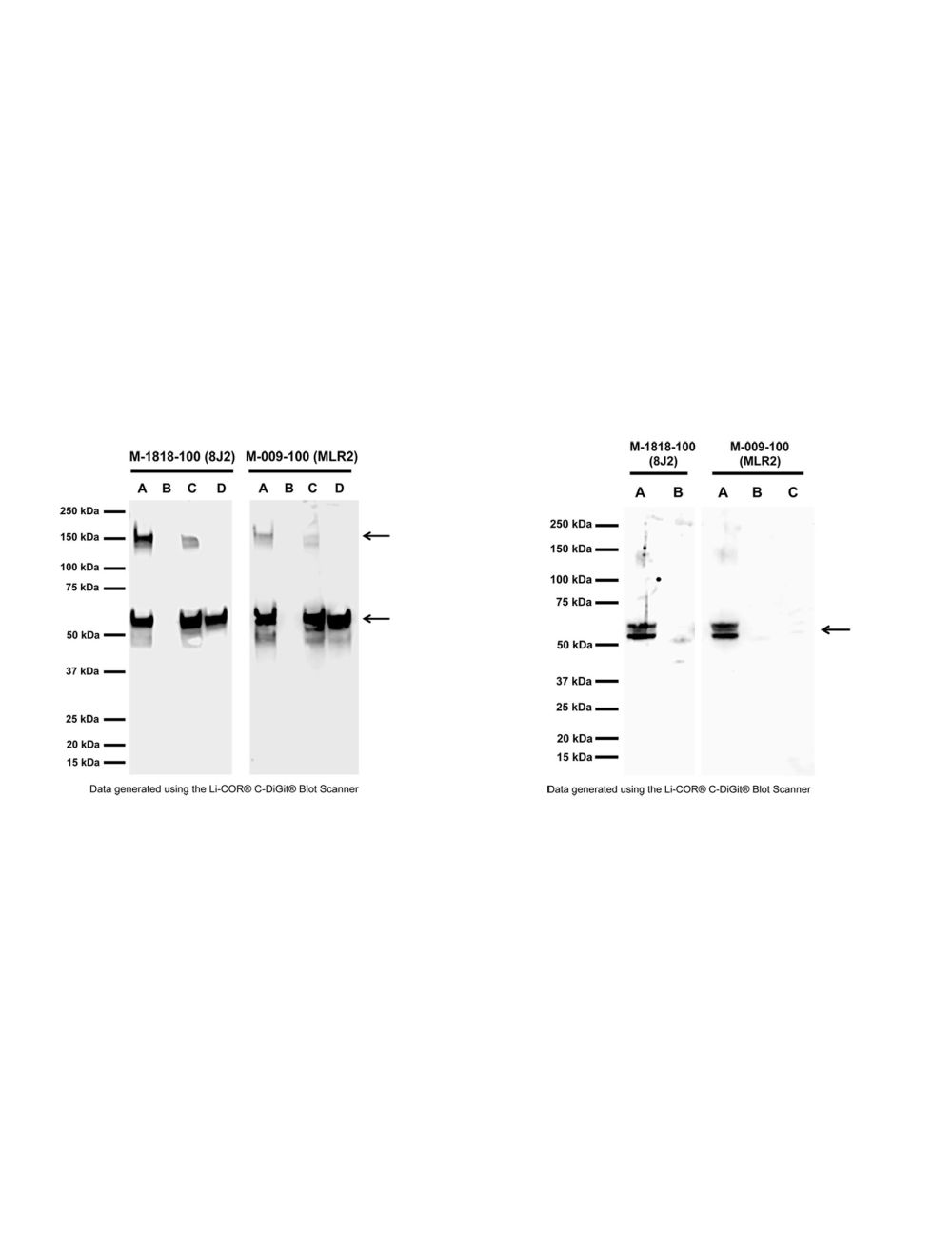

NGFR/p75 neurotrophin receptor (p75NTR), Mouse Monoclonal Antibody
- Product Name NGFR/p75 neurotrophin receptor (p75NTR), Mouse Monoclonal Antibody
-
Product Description
Mouse anti-p75 neurotrophin receptor (p75NTR) Monoclonal Antibody (Unconjugated), suitable for FC, ICC, IHC-Frozen, WB, IP, Immunopanning.
- Alternative Names Low-affinity nerve growth factor receptor; NGF receptor; Gp80-LNGFR; p75 ICD; Low affinity neurotrophin receptor p75NTR; CD271
- Application(s) FC, ICC, IHC-Frozen, Immunopanning, IP, WB
- Antibody Host Mouse
- Antibody Type Monoclonal
- Specificity Human, reacts with human, mouse and rat. Cross-reactivity with other species not tested but expected.This antibody is specific for NGFR/p75NTR as demonstRated by western blotting and immunprecipitation. The antibody recognizes extracellular p75NTR under non-reducing conditions.
- Species Reactivity Human, Mouse, Rat
- Immunogen Description Recombinant extracellular domain (amino acids 29-250) of human NGFR/p75NTR protein with N-terminal His-tag.
- Conjugate Unconjugated
- Purity Description Protein A purified IgG
- Regulatory Status For research use only.
Product Info
-
Product Description
Mouse anti-p75 neurotrophin receptor (p75NTR) Monoclonal Antibody (Unconjugated), suitable for FC, ICC, IHC-Frozen, WB, IP, Immunopanning.
-
Related Products
NGFR/p75 neurotrophin receptor (p75NTR), Mouse Monoclonal Antibody (FITC)
NGFR/p75 neurotrophin receptor (p75NTR), Mouse Monoclonal Antibody (ATTO 488)
- Application(s) FC, ICC, IHC-Frozen, Immunopanning, IP, WB
-
Application Details
Flow Cytometry: 5-20 µg/mL.
Western Blotting: 0.5-2.0 µg/mL, non-reducing conditions only (no DTT or beta-mercaptoethanol).
Immunoprecipitation: lysate dependent. 10 ug per 200-500 ug total protein.
Immunopanning: 1-5 µg/mL.
Immunocytochemistry: 1-5 µg/mL. Staining is strongest in non-fixed cells, light fixation is tolerable.
Immunohistochemistry: fresh, acetone fixed sections only, epitope is fixation sensitive. Not suitable in formalin-fixed, paraffin (FFPE) embedded tissues.
Biosensis recommends optimal dilutions/concentrations should be determined by the end user. - Target NGFR/p75 neurotrophin receptor (p75NTR)
- Specificity Human, reacts with human, mouse and rat. Cross-reactivity with other species not tested but expected.This antibody is specific for NGFR/p75NTR as demonstRated by western blotting and immunprecipitation. The antibody recognizes extracellular p75NTR under non-reducing conditions.
- Target Host Species Human
- Species Reactivity Human, Mouse, Rat
- Antibody Host Mouse
- Antibody Type Monoclonal
- Antibody Isotype IgG2a
- Clone Name 8J2
- Conjugate Unconjugated
- Immunogen Description Recombinant extracellular domain (amino acids 29-250) of human NGFR/p75NTR protein with N-terminal His-tag.
- Purity Description Protein A purified IgG
- Format Lyophilized from a solution containing PBS buffer pH 7.2-7.6 with 0.1% trehalose, without preservatives.
- Reconstitution Instructions Spin vial briefly before opening. Reconstitute in 100 µL sterile-filtered, ultrapure water to achieve a concentration of 1 mg/mL. Centrifuge to remove any insoluble material. Final buffer contains no preservatives.
- Storage Instructions Store lyophilized antibody at 2-8°C. After reconstitution divide into aliquots and store at -20°C for long-term storage. Store at 2-8°C short-term (up to 4 weeks) with an appropriate antibacterial agent. Avoid repetitive freeze/thaw cycles.
- Batch Number Please see item label.
- Expiration Date 12 months after date of receipt (unopened vial).
- Alternative Names Low-affinity nerve growth factor receptor; NGF receptor; Gp80-LNGFR; p75 ICD; Low affinity neurotrophin receptor p75NTR; CD271
- Uniprot Number P08138
- Uniprot Number/Name P08138 (TNR16_HUMAN)
- Scientific Background p75NTR (CD271) was originally discovered as a low affinity nerve growth factor receptor (NGFR). Later it was found that it was the receptor for all neurotrophins, including NGF, BDNF, NT3 and NT4/5. It mediates signals of neurotrophins for neuronal survival, apoptosis, neurite outgrowth and synaptic plasticity. Recently, it has been revealed that p75NTR not only acts as the receptor for neurotrophins but also the receptor for many other pathological ligands such as prions, rabies virus and amyloid beta. p75NTR also acts as a co-receptor for NOGO which mediates inhibitory signals of myelin associated protein. p75NTR is highly expressed in a number of non-neuronal and neuronal cells including motor neurons during development and also in damaged neurons. Recent research proposes the extracellular domain of p75NTR as a biomarker for monitoring the progression of motor neuron disease (MND), also known as Amyotrophic Lateral Sclerosis (ALS) or Lou Gehrig's Disease. SUBUNIT: Homodimer; disulfide-linked. Interacts with p75NTR-associated cell death executor. Interacts with NGFRAP1/BEX3.
- Shipping Temperature 25°C (ambient)
- UNSPSC CODE 41116161
- Regulatory Status For research use only.
Specifications


-
Specific References
Salehi S et al. (2023) Cytosolic Ptbp2 modulates axon growth in motoneurons through axonal localization and translation of Hnrnpr Nat Commun. 14(1):4158. Application: Mouse, Immunopanning.
Hennlein L et al. (2023) Plastin 3 rescues cell surface translocation and activation of TrkB in spinal muscular atrophy J Cell Biol. [Epub ahead of print] Application: Mouse, Immunopanning.
Hulme AJ et al. (2020) Molecular and Functional Characterization of Neurogenin-2 Induced Human Sensory Neurons . Front Cell Neurosci. 14:600895 Application: Human, ICC(IF). -
General References
Matusica D et al. (2008). Characterisation and use of the NSC-34 cell liner for study of nerurotrophin receptor trafficking. J. Neurosci. Res. 86(3) pp. 553-65.
Huh CY et al. (2008). Chronic exposure to nerve growth factor increases acetylcholine and glutamate release from cholinergic neurons of the rat medial septum and diagonal band of Boca via mechanisms mediated bu p75NTR. J. Neurosci. 28(6) pp. 1404-9.
Lagares A et al. (2007). Primary sensory neuron addition in the adult rat trigeminal ganglion: evidence for neural crest glio-neuronal precursor maturation. J. Neurosci. 27(30) pp. 7939-53.
DiStefano & Johnson (1988). Identification of a truncated form of the nerve growth factor receptor. Proc Natl Acad Sci U S A. 1988 Jan;85(1):270-4.